


Recent News
Assessing Kidney Disease Globally
Kidney disease worsens outcomes for patients hospitalized with cirrhosis; study finds.
Read MoreAssessing Kidney Disease Globally
Kidney disease worsens outcomes for patients hospitalized with cirrhosis; study finds.
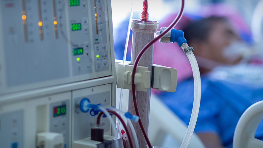
Image Caption: Chronic kidney disease, a progressive condition in which the kidneys lose their ability to function properly, is a growing global health concern. It can progress to end-stage kidney failure requiring dialysis or kidney transplant.
The global prevalence of chronic kidney disease (CKD) is increasing, leading to higher mortality among affected individuals and placing a growing burden on health care systems. A large international study co-led by UHN examined the prevalence and impact of CKD in those with cirrhosis—permanent scarring and damage of the liver. The researchers found that CKD negatively impacts the prognosis of patients with cirrhosis in a global cohort.
CKD occurs when the kidneys are damaged or do not function as they should for a prolonged period of time. It was once thought to be uncommon in patients with cirrhosis. However, its prevalence has increased among those with advanced liver disease. Experts say this increase is being driven by growing rates of other co-occurring metabolic conditions such as obesity, diabetes, and high blood pressure, as well as broader diagnostic criteria that identify CKD earlier.
However, most evidence on CKD in cirrhosis comes from Western countries, with minimal reporting from Africa, South America, or Oceania. To assess the global prevalence of CKD in cirrhosis and its impact on patient outcomes, the research team analyzed data from more than 7,000 hospitalized patients with cirrhosis enrolled in the Chronic Liver Disease Evolution and Registry for Events and Decompensation (CLEARED) consortium.
The study found that approximately 18% of people hospitalized with cirrhosis also had CKD. Rates were highest in high-income countries, where metabolic syndrome—a cluster of conditions that includes obesity, diabetes, high blood pressure, high cholesterol and fatty liver disease—is more common.
In patients with cirrhosis, CKD was more likely seen in those with ascites, a liver complication caused by a build-up of fluid in the abdomen. Patients with both CKD and cirrhosis also had more complex liver complications. Acute kidney injury (AKI), a sudden decline in kidney function, was the most common complication during hospitalization. It affected nearly 60% of patients with CKD, more than double the rate seen in patients without CKD. Patients in the hospital with both CKD and cirrhosis also had a higher risk of death during or shortly after hospitalization.
The results show that CKD negatively impacts outcomes for patients with cirrhosis, highlighting the need for closer monitoring and treatment of kidney disease in these patients. Careful management of ascites and lifestyle changes aimed at reducing risk factors could help improve outcomes, particularly in high-income countries.
Dr. Florence Wong is an Affiliate Scientist at UHN’s Organ Systems and Integrated Health Sciences Research Institute and a Professor in the Department of Medicine at The University of Toronto. She is the lead and corresponding author of the study.
This research was supported by UHN Foundation.
Wong F, Adebayo D, George J, Idilman R, Hayes PC, Alvares-da-Silva MR, Torre A, Mekonnen HD, Seto WK, Sarin SK, Cao Z, Rajoriya N, Nagral A, Fisseha H, Kulkarni AV, Zhu C, Debzi N, Farias AQ, Su M, Goel A, Marciano S, Livingstone R, Dhiman RK, Gao Y, Malé-Velázquez R, Demitars CO, Bera C, Jiang YF, Velarde-Ruiz Velasco JA, Tan HK, Zhao C, Huezo MSG, Asrani SK, Wang L, Lu M, Michalczuk MT, Barutcu S, Cordova-Gallardo J, Gofton C, Gounder M, Shaw J, Albhaisi S, Zheng X, Aravinthan A, Rajaram RB, Jothimani D, Thanapirom K, Benitez C, Dincer D, Saraya A, Xu B, Lin M, Wu X, Liu C, Reddy R, Bush B, Thacker LR, Topazian M, Xie Q, Silvey S, Kamath PS, Choudhury A, Bajaj JS; CLEARED Investigators. Chronic kidney disease in cirrhosis: a study of inpatients from a global perspective. Gut. 2026 Jun 5. doi: 10.1136/gutjnl-2025-336802. Epub ahead of print.
×

Creating Roadmaps to Better Care
Study explores what patients and caregivers identify as priorities for improved hospital stays.
Read MoreCreating Roadmaps to Better Care
Study explores what patients and caregivers identify as priorities for improved hospital stays.
Image Caption: Patient and caregiver input can help guide improvements to the quality of care in general medicine wards, potentially enhancing the experiences and health outcomes of many patients.
General medical wards in hospitals provide care to large and growing numbers of patients. As a result, identifying effective ways to improve the quality of care can enhance the experiences and outcomes of many individuals. A new study from UHN and the University of Toronto is shedding light on what matters most to patients and caregivers during a hospital stay, revealing that better communication, fewer delays, and greater inclusion in care decisions are among the top priorities for improvement.
By using an open-ended, multi-step approach, known as group concept mapping, researchers led by Dr. Lauren Lapointe-Shaw, Staff Physician in General Internal Medicine and Scientist at UHN’s Organ Systems & Integrated Health Sciences Research Institute, sought to identify high-priority areas directly from patients' and caregivers' experiences.
Over 500 patients and caregivers from general medical wards at 10 Canadian hospitals (in Ontario, Nova Scotia, British Columbia, and Alberta) participated in this multiphase study between November 2023 and February 2025. Participants included people from groups that are often underrepresented in health research, such as non-English speakers, people experiencing homelessness, individuals with mental health conditions, and people living with dementia.
Patients and caregivers first told researchers what contributed to their inpatient experience, and then later rated and sorted the resulting experience statements. The sorting exercise resulted in six main themes: Environment and Facilities, Personal Support, Delays, Communication, Nutrition, and Inclusivity and Engagement.
Overall, patients’ and caregivers’ own negative inpatient experiences, many of them related to the hospital environment, did not correlate with what issues they prioritized for improvement. Instead, it was system-level concerns such as care delays, communication breakdowns, and lack of inclusivity that were seen as the most important to improve. One example of this is how rare but harmful events, such as perceived discrimination, were prioritized for improvement by many, despite few having experienced these directly. Notably, priorities for improvement were consistent across different patient and caregiver groups, even when their experiences varied.
Although negative experience ratings and priorities for improvement were not strongly correlated, the top-rated item was the same for both: having to wait a long time in the emergency department before getting a bed on the ward. This issue negatively affected many patients and was also identified as the highest priority for improvement.
The other highest-ranked priorities for improvement centered around communication, inclusivity, and engagement. For example, not having enough opportunities to speak with a doctor was another top priority for improvement.
“This study offers a patient-defined roadmap to guide quality improvement efforts on inpatient wards,” says Dr. Lapointe-Shaw. “Our findings also demonstrate that patients and caregivers can and do make a distinction between their own negative experiences and what they consider most essential to improve, for the benefit of all patients.”
Dr. Lauren Lapointe-Shaw is a Scientist at UHN’s Organ Systems & Integrated Health Sciences Research Institute, Research Director of the Myrna Daniels Seniors Emergency Medicine Centre at UHN, and Associate Professor in the Departments of Medicine & Institute of Health Policy, Management and Evaluation (IHPME) at the University of Toronto. She is the lead and senior author of the study.
This work was supported by the Canadian Institutes of Health Research, the PSI Foundation, the Sinai UHN Medical Organization, St Michael’s Hospital Academic Medical Organization, and UHN Foundation.
Lapointe-Shaw L, Salahub C, Lee M, Verma AA, Razak F, Ambasta A, Refaei M, Carpenter T, Moses B, Shetty N, Miller AP, Spicer E, Rawal S, Soong C, Kuluski K, Okrainec K, Baker GR, Chartier LB, Dhalla IA, Joseph R, Wodchis WP, Desveaux L, Wu W, Granieri M; GIM Priorities Group. Patient and caregiver priorities for quality improvement on general medical wards: a multicentre group concept mapping study. BMJ Qual Saf. 2026 Jul 19:bmjqs-2026-020280. doi: 10.1136/bmjqs-2026-020280. Epub ahead of print.
×

Finding the Right Balance
Repeated reactive balance training was associated with better balance control after stroke.
Read MoreFinding the Right Balance
Repeated reactive balance training was associated with better balance control after stroke.
Image Caption: Stroke can affect balance responses needed for safe movements, increasing fall risk and affecting quality of life. Understanding how rehabilitation can help restore these critical skills may help improve independence and mobility after stroke.
For people recovering from a stroke, regaining the ability to walk independently is essential for returning to daily life. But safe mobility depends on being able to quickly recover from a stumble or sudden loss of balance—an ability that is often impaired after stroke. New research from UHN’s KITE Research Institute suggests that increased exposure to reactive balance training improves balance control after stroke.
Reactive balance training is a rehabilitation approach that intentionally exposes participants to controlled balance disturbances to help them practice regaining their balance in a safe environment. In previous studies, this training approach has shown promise in improving balance control after stroke; however, clinicians remain uncertain about how much training is needed to achieve meaningful improvements and which training characteristics matter most.
To explore this, the research team studied 30 people recovering from stroke who completed up to 12 one-hour training sessions over six weeks. During the sessions, participants practised recovering their balance from controlled balance disturbances triggered by activities such as kicking a soccer ball, walking on the spot, or responding to controlled pushes. Researchers tracked the number of balance disturbances experienced, task difficulty, success rate, and participants’ perceived level of challenge.
The findings showed that participants who experienced a greater number of balance disturbances showed larger improvements in reactive balance control. In contrast, task difficulty, success rate, and the perceived challenge level were not associated with better outcomes. The results suggest that repeated exposure to balance-recovery practice may play an important role in improving reactive balance control after stroke.
By emphasizing training volume and consistent exposure to balance challenges, clinicians may be able to better tailor rehabilitation programs and improve recovery outcomes for people living with the long-term effects of stroke. Further studies should explore how different task types interact with different individuals.
Júlia O. Faria, first author of the study, was a visiting PhD student from the University of São Paulo, Brazil.
Dr. Avril Mansfield, senior author of the study, is a Senior Scientist at UHN’s KITE Research Institute. At the University of Toronto, she is an Associate Professor in the Department of Physical Therapy and a Faculty Member at the Rehabilitation Sciences Institute.
This work was supported by UHN Foundation, the Canadian Institutes of Health Research, Canada Foundation for Innovation, the Ontario Government, and CAPES (Coordination for the Improvement of Higher Education Personnel).
Faria JO, Danells CJ, Inness EL, Mansfield A. Optimal Reactive Balance Training Characteristics Poststroke: Secondary Analysis of a Clinical Trial. Physiother Res Int. 2026 Jul. doi: 10.1002/pri.70231.
×
Global Talent, Canada’s Future
New federal funding to support 13 international researchers advancing Canadian innovation.
Read MoreGlobal Talent, Canada’s Future
New federal funding to support 13 international researchers advancing Canadian innovation.
UHN has secured a total of $1.7 million to recruit 13 internationally-based doctoral and postdoctoral researchers through Phase 1 of the Government of Canada’s Canada Impact+ Research Training Awards.
These awards are part of the Canada Global Impact+ Research Talent Initiative, a $1.7 billion federal investment to recruit over 1000 researchers to Canada and support their continued contributions to Canadian innovation. The Research Training Awards support doctoral scholarships and postdoctoral research awards across eight priority research areas, including health and advanced digital technologies like AI.
Through this funding, UHN will welcome 8 doctoral students and 5 postdoctoral researchers. Their work will focus on major disease areas, including cancer, immune-related conditions, and heart, lung, and liver diseases. Using approaches such as AI-enabled diagnostics, stem cell and regenerative medicine, and immune-based therapies, these researchers will improve patient care and outcomes.
These results reflect the strength of UHN’s research community and support institutional efforts to attract top international talent, such as UHN’s Canada Leads program.
Congratulations and welcome to the newest members of TeamUHN. Read the full press release here.
×
New Clues to Leukemia Relapse
Researchers uncover how DNA elements may fuel leukemia recurrence and therapy resistance.
Read MoreNew Clues to Leukemia Relapse
Researchers uncover how DNA elements may fuel leukemia recurrence and therapy resistance.
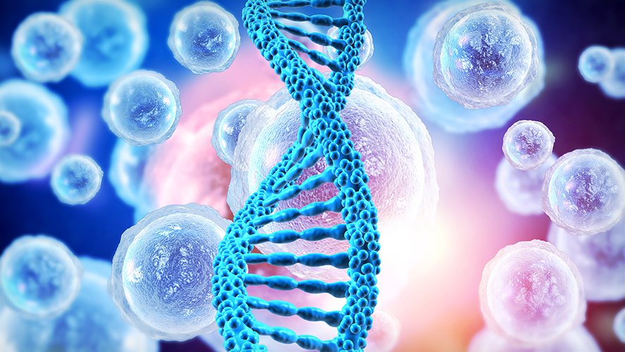
Image Caption: Leukemia stem cells, which can self-renew and produce more cancer cells, are thought to be responsible for disease progression and relapse. Scientists have found that the genetic features of leukemia stem cells can help predict disease outcomes and identify patients at higher risk of relapse.
A new study from UHN’s Princess Margaret Cancer Centre (PM) found that ancient DNA sequences in our genome, called transposable elements, can act as control hubs for leukemia stem cells, fueling therapy resistance and relapse in acute myeloid leukemia (AML). Although most patients with AML achieve complete remission after chemotherapy, two-thirds will relapse within five years.
Lifelong blood production originates from self-renewing, hematopoietic stem cells (HSCs)—cells that give rise to different types of immature (precursor) and mature blood cells. In blood cancers such as leukemia, cancer stem cells can self-renew and give rise to cancerous cells.
A small group of powerful cancer stem cells, called leukemia stem cells (LSCs), is one of the biggest obstacles to curing blood cancers. LSCs can survive chemotherapy, reignite the disease, and drive relapse. However, the genetic reasons why LSCs remain so resilient are poorly understood.
By comparing how DNA is packaged and accessed (a process known as epigenetics) in LSCs, HSCs, and more mature blood cells, researchers found that transposable elements—sometimes called "jumping genes" because they can move around the genome—help distinguish stem cells from more mature, specialized blood cells.
The team, including first authors Drs. Giacomo Grillo and Bettina Nadorp, found that specific families of transposable elements, such as ERV1 and ERV3, are highly active in LSCs. These same elements normally function in healthy blood stem cells to support self-renewal.
Transposable elements are an under-explored class of DNA sequences, making up roughly 50% of the human genome. “They are not genes, but they can turn on genetic programs that allow cells to renew and thrive. In this way, leukemia stem cells hijack what healthy blood stem cells use for regeneration, fueling cancer relapse instead,” says Dr. Mathieu Lupien, Senior Scientist at PM and co-senior author of the study.
Building on this discovery, the researchers developed a genomic score that can identify patients with AML who are more likely to relapse. The score measures how closely a patient's leukemia cells resemble LSCs by examining whether specific families of transposable elements are found in open, accessible regions of the genome.
Through efforts led by Dr. John Dick, Senior Scientist at PM, and his team, including Senior Scientific Associate Dr. Helena Boutzen, chemical tools were used to selectively mask and disable certain transposable elements in preclinical models of leukemia. Doing so resulted in LSCs losing their ability to renew and thrive, opening the door to new possible therapeutic strategies.
“Together, these findings give us hope that we can eliminate leukemia at its root and not just decrease the burden of the disease,” says Dr. Lupien. “We may finally change outcomes for patients living with AML.”
Dr. Giacomo Grillo, former postdoctoral researcher at UHN and currently a Staff Scientist at the European Institute of Oncology, is a co-first author of the study.
Dr. Bettina Nadorp, former postdoctoral researcher at UHN and currently an Associate Director at Bristol Myers Squibb, is a co-first author of the study.
Dr. Helena Boutzen, Senior Scientific Associate at Princess Margaret Cancer Centre, is a co-author of the study.
Dr. John Dick, Senior Scientist at Princess Margaret Cancer Centre, University Professor at the University of Toronto, Helga and Antonio De Gasperis Chair in Blood Cancer Stem Cell Research, and Professor in the Department of Molecular Genetics at the University of Toronto, is a co-corresponding author of the study.
Dr. Mathieu Lupien, Senior Scientist at Princess Margaret Cancer Centre and Professor in the Department of Medical Biophysics at the University of Toronto, is a co-corresponding author of the study.
This work was supported by The Princess Margaret Cancer Foundation, Ontario Institute for Cancer Research, the Province of Ontario, the Canadian Institutes for Health Research (CIHR), Medicine by Design, The Terry Fox Research Institute, the Canada Research Chairs program, the Canadian Cancer Society Research. Dr. Lupien holds the Joey and Toby Tanenbaum/Brazilian Ball Chair in Prostate Cancer Research at UHN's Princess Margaret Cancer Centre.
Giacomo Grillo, Bettina Nadorp, and Aditi Qamra report that at the time of publication, they were employees of T-One Therapeutics, Bristol Myers Squibb, and Hoffman-La Roche Canada, respectively.
Grillo G, Nadorp B, Qamra A, Drylie B, Mitchell A, Arlidge C, Nand A, Takayama N, Murison A, Madani Tonekaboni SA, Kang KK, Arruda A, Wang JCY, Minden MD, Deniz Ö, Boutzen H, Dick JE, Lupien M. Transposable elements shape stemness in normal and leukemic hematopoiesis. Nat Genet. 2026 May. doi: 10.1038/s41588-026-02585-z. Epub 2026 May 4.
×

Guiding Equal Heart Failure Care
Study finds new strategy for heart failure care is equally effective for males and females.
Read MoreGuiding Equal Heart Failure Care
Study finds new strategy for heart failure care is equally effective for males and females.
Image Caption: Heart failure can look different in male and female patients, with different underlying factors, symptom burden, and hospitalization rates. Therefore, it is important to determine whether emergency department approaches to acute heart failure management are effective for these different patient populations.
Heart failure is one of the leading causes of illness and mortality worldwide. The care strategy used by emergency departments to manage heart failure can significantly impact patient outcomes. A recent UHN-led study found that a new strategy used to manage patients with this condition is equally effective for male and female populations.
When patients come to the emergency room with heart failure, it can be difficult to determine whether they need to be admitted or can safely be discharched. Discharging high-risk patients too early can lead to serious complications or death. A major Canadian trial (the COACH trial) co-led by UHN’s Dr. Douglas Lee showed that using a structured risk assessment tool and providing rapid follow-up care after discharge can improve outcomes—but it was unclear whether these benefits were the same for males and females.
Although male and female patients have a similar lifetime risk of developing heart failure, female patients often face a higher burden of symptoms, lower quality of life, and more hospital readmissions despite lower rates of hospitalization overall. Some research has also suggested that females may be more likely than males to be readmitted to hospital for heart failure within a year of discharge.
Because of this disparity in outcomes with the current standard of care, researchers wanted to know whether the COACH trial strategy works equally well for both males and females.
Researchers analyzed data from more than 5,400 patients treated across 10 hospitals in Ontario between 2017 and 2019 as part of the COACH clinical trial.
After a follow-up period of both 30 days and 20 months, both male and female patients saw similar outcomes. For both patient groups, the strategy in the COACH trial led to modest reductions in the combined risk of death or heart-related hospitalization, with no meaningful difference between sexes.
Overall, the findings suggest that using risk-based decision tools in the emergency department, combined with coordinated follow-up care, can benefit heart failure patients regardless of sex. These results support the application of this care model broadly, offering more patients with heart failure improved outcomes.
Dr. Douglas Lee is the Division Head of Cardiology and a Senior Scientist at UHN’s Peter Munk Cardiac Centre, and a Professor in the Department of Medicine and the Institute of Health Policy, Management and Evaluation at the University of Toronto. He is also a Senior Core Scientist at ICES and the Ted Rogers Chair in Heart Function Outcomes at the Ted Rogers Centre for Heart Health. He is the first and corresponding author of the study.
Dr. Heather Ross is a Clinician Scientist at UHN's Peter Munk Cardiac Centre, the Loretta Rogers Chair in Heart Function at UHN, and a Professor of Medicine at the University of Toronto. She is a co-senior author of the study.
This work was supported by the Ontario SPOR Support Unit, the Ted Rogers Centre for Heart Research, the Canadian Institutes of Health Research, ICES, the Ontario Ministry of Health (MOH) and Ministry of Long-Term Care, and UHN Foundation.
For a list of competing interests, see the manuscript.
Lee DS, Wang CN, Austin PC, Straus SE, Farkouh ME, Chong A, Taljaard M, Poon S, Smith S, McKelvie RS, Iwanochko RM, MacDougall A, Elbarasi E, Cram PM, Fang J, Atzema CL, Udell JA, Rochon PA, Schull MJ, Mak S, Ross HJ. Risk-Stratified Transitional Care and Cardiovascular Hospitalizations by Sex: A Secondary Analysis of a Randomized Clinical Trial.JAMA Netw Open. 2026 May 1. doi: 10.1001/jamanetworkopen.2026.11892.
×

Empowering Fair and Responsible AI
A team led by Princess Margaret developed AFFRM-AI, a framework for AI in clinical settings.
Read MoreEmpowering Fair and Responsible AI
A team led by Princess Margaret developed AFFRM-AI, a framework for AI in clinical settings.
Image Caption: AFFRM-AI is designed to guide research teams and clinicians on how to responsibly, safely, and compassionately develop and deploy AI solutions for health care.
Health care is increasingly being transformed by AI, with technologies such as medical scan analysis and cancer risk prediction tools enabling faster diagnosis and treatment. Yet, alongside these advancements comes a critical challenge: how do we translate powerful AI methods into safe, trustworthy, and sustainable clinical tools? To address this, UHN’s Cancer Digital Intelligence (CDI), a research and innovation program within the Princess Margaret Cancer Centre, developed AFFRM-AI, a framework for the fair and responsible use of AI tools.
Bridging a Critical Gap: AFFRM-AI
CDI aims to improve cancer care through technology and AI. As such, it has been actively researching the use of AI in medicine since the technology’s early development. One goal of CDI is to promote a culture of fair and responsible AI and take the steps necessary to mitigate bias in AI solutions caused by social, data-related, methodological or algorithmic factors. Despite the growth of AI use, there has been a lack of accessible and actionable resources on the fair and safe use of AI in clinical settings.
To address this, CDI assembled a 16-member working group composed of leading experts from various institutions in related fields including AI, bias, ethics, privacy, education and medicine to develop a guidance document.
The CDI team developed, A Framework for Fair and Responsible Machine Learning and AI (AFFRM-AI) that outlines best practices and recommendations for two key goals:
- Encouraging responsible, safe, and compassionate AI development and deployment within UHN clinics
- Safeguarding patients and care providers from biased and inequitable AI predictions.
AFFRM-AI provides actionable guidance across four key stages: (1) problem identification and study design, (2) model development and training, (3) silent deployment—testing real-world performance observationally without influencing clinical decisions—and clinical evaluation, and (4) operational deployment and ongoing monitoring. Within each stage, the framework provides concrete recommendations, reflective questions, and documentation prompts addressing how data are used and approved, data readiness and availability, how fairness is measured, selection of appropriate outcome measures, and more.
Designed to adapt to local contexts, AFFRM-AI enables multidisciplinary teams—including clinicians, data scientists, privacy experts, ethicists, and operational leaders—to share a common language and set clear expectations for building safe AI systems that are grounded in equity, fairness, scientific rigor, and transparency.
Driving Responsible AI at UHN and Beyond
The AFFRM-AI framework and the methodology behind its development have been published in The Lancet Digital Health. The team is now focused on cultivating engagement within the UHN community, including working with the Research Ethics Board (REB) and the AI Deployment team, among others, to encourage organization-wide adoption of standards for equitable and responsible AI use.
By providing concrete, operational guidance, AFFRM-AI aims to put responsible AI into practice and protect patients. Initiatives such as this position UHN at the forefront of responsible innovation, ensuring that powerful AI methods and progress translate into safe, trustworthy, and sustainable clinical tools.
For more information and to view the Framework, see here.
The AFFRM- AI Framework was prepared by Mattea Welch, Benjamin Grant, and Christopher Deutschman, in consultation with Clare McElcheran, Adam Badzynski, Jennifer A.H. Bell, Andrew Hope, Robert C. Grant, Tran Truong, Kelly Lane, Patti Leake, Divya Sharma, Ian Stedman, Mike Lovas, Ale Berlin, Jeremy Petch, Benjamin Haibe-Kains, and James A. Anderson.
Work on AFFRM-AI was partly supported by the Associated Medical Services (AMS) Healthcare, CDI and The Princess Margaret Cancer Foundation.
See the manuscript for additional acknowledgements and competing interests.
Welch ML, Grant B, Deutschman C, McElcheran C, Badzynski A, Bell JAH, Hope A, Grant RC, Truong T, Lane K, Leake P, Sharma D, Stedman I, Lovas M, Petch J, Berlin A, Haibe-Kains B, Anderson JA. A practical framework for operationalising responsible and equitable artificial intelligence in health care: tackling bias, inequity, and implementation challenges. Lancet Digit Health. 2026 Mar;8(3):100957. doi: 10.1016/j.landig.2025.100957. Epub 2026 Mar 20.
×
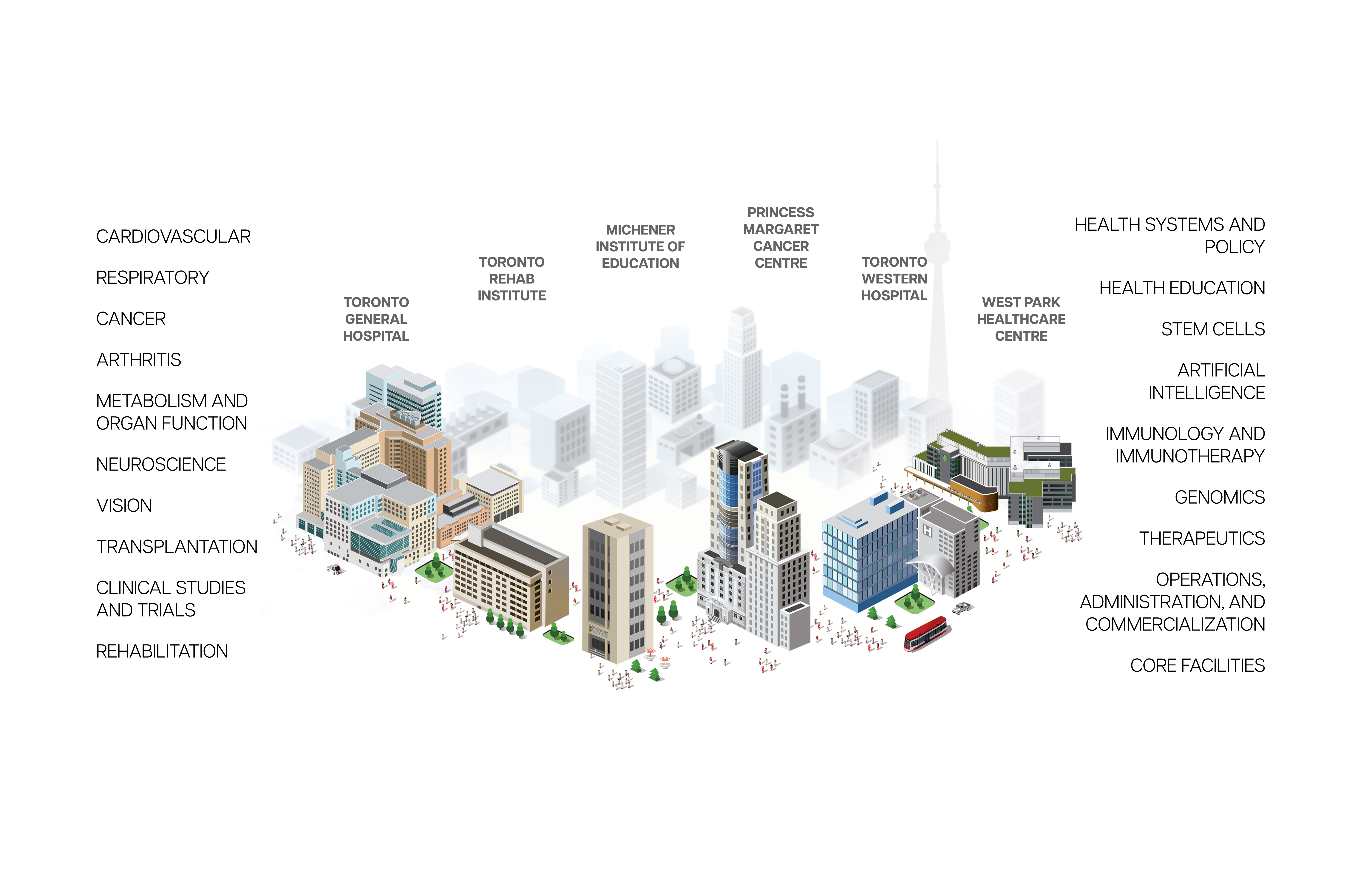
Research Institutes at UHN
Research conducted at UHN's research institutes spans the full spectrum of diseases and disciplines, including cancer, cardiovascular sciences, transplantation, neural and sensory sciences, musculoskeletal health, rehabilitation sciences, and community and population health.
Research Institutes
Research at UHN is conducted under the umbrella of the following research institutes. Click below to learn more: