


Biological tissues, like those in the kidney, are often organized into complex patterns, which are an integral part of how they function. A new study from scientists at UHN found that incorporating specific, intricate patterns in lab-grown kidney tissue can enhance how these models function and are used in research.
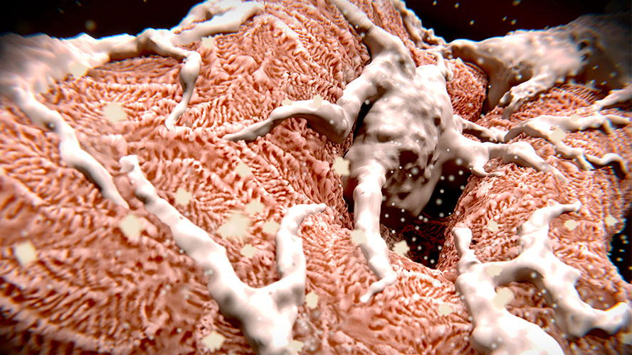
Kidneys filter the blood—removing waste and balancing fluids. Within the kidney, this filtering is done through networks of looping capillaries (tiny blood vessels), called glomeruli. Specialized cells called podocytes then wrap around these blood vessels using cellular projections called foot processes that branch from the main cell.
Podocytes provide a key filtration barrier. As they develop, podocytes' foot processes become increasingly complex. This complexity is an important indicator of podocyte health and function. Natural podocyte branching appears to follow fractal patterning—a form of repetition in which successively smaller copies of a pattern are nested inside each other, like tree branches.
These fractal patterns are not typically recreated in standard laboratory cell cultures—cells grown in a controlled laboratory environment that can be used as tools to test biological processes. Despite recent advancements, lab-grown models fail to replicate the complex 3D architecture of kidney tissue. This leads to models that are less functional and mature than those in the body, potentially hindering their use in the study of kidney function and disease.
As a proper 3D shape is known to influence podocyte growth and function, the research team explored whether incorporating a realistic architecture with fractal patterning in cell cultures could make lab models better and more representative of real kidneys.
First, by comparing detailed images of kidney cells, including podocytes, to fractal and non‑fractal shapes, they confirmed that healthy podocytes do indeed form fractal patterns in nature. They then created artificial textured surfaces that mimic these natural fractal patterns to grow cells on in the lab.
The team found that podocytes grown on these fractal surfaces showed stronger markers of functionality, had enhanced maturation, and organized themselves more like they do in the body compared to those grown on flat surfaces. Gene analysis also showed that these cells produced more of the proteins that make up the supportive material around them.
Overall, the results suggest that this approach to growing cells could eventually improve lab kidney models—advancing research in kidney disease and treatments.
Chuan Liu is a Doctoral student in Dr. Radisic’s lab and first author of the study.
Dr. Milica Radisic is a Senior Scientist at UHN and a Professor at the Institute of Biomedical Engineering at the University of Toronto. She is the corresponding author of the study.
This work was supported by the Natural Sciences and Engineering Research Council of Canada (NSERC), the Canadian Institutes of Health Research (CIHR), the Center for Research and Applications in Fluidic Technologies, the Canada Foundation for Innovation, Additional Ventures, and UHN Foundation.
Dr. Milica Radisic is a Tier 1 Canada Research Chair in Organ-on-a-Chip Engineering.
Drs. Lieu, Konvalinka and Radisc are inventors on a pending US Patent application relating to fractal cues for cell maturation.
Liu C, Aggarwal P, Wagner KT, Landau S, Cui T, Song X, Shamaei L, Rafatian N, Zhao Y, Rodriguez-Ramirez S, Morton K, Virlee E, Li CY, Bannerman D, Pascual-Gil S, Okhovatian S, Radisic A, Clotet-Freixas S, Veres T, Sadrzadeh M, Filleter T, Broeckel U, Konvalinka A, Radisic M. Biomimetic fractal topography enhances podocyte maturation in vitro. Nat Commun. 2025 Dec 15;16(1):11116. doi: 10.1038/s41467-025-66037-8.