A Bridge Beyond Pacemakers
UHN team generates cardiac-like cells that could replace electronic pacemakers after AV block.
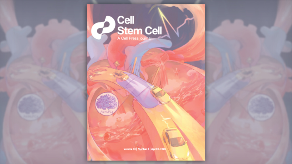
The cover image of Cell Stem Cell depicts the heart’s conduction system as a highway, with cars symbolizing electrical signals. The cars are crossing a damaged AVN bridge representing AV block. A new innovation using heart cells grown in the lab acts as a bandage to restore this passage. Illustration by Maggie Kwan; Cover Cell Stem Cell ©2026.
A research team from UHN’s McEwen Stem Cell Institute (McEwen) has grown heart cells in the lab that mimic the atrioventricular node (AVN), the part of the heart that carries electrical impulses from the atria to the ventricles. This innovation could offer individuals with a dysfunctional AVN a novel treatment option—called a biological conduction bridge—that may one day replace standard pacemaker implants.
As the “electrical bridge” between the atria and ventricles, the AVN is responsible for maintaining the pace of the ventricles’ contractions and, thus, efficient blood flow. An improperly functioning AVN can lead to a life-threatening condition called atrioventricular (AV) block, which is typically treated by implanting an electronic pacemaker (EPM) to take over the AVN’s function. Although lifesaving, EPMs are not without limitations, including the need for surgery to replace their batteries every 10 to 15 years and their inability to respond to physiological changes. These limitations underscore the need for alternative approaches that more closely mimic native cardiac conduction.
In pursuit of an alternative, cell-based therapy for AV block, Dr. Stephanie Protze, a McEwen Scientist, and her team used a specialized lab process that turns stem cells into specific cell types to create cardiac pacemaker cells that behave like those in the AVN (AVNLPCs). The team then assessed gene expression and functional properties to confirm the cells’ identity as AVNLPCs.
The AVNLPCs showed the same key biological markers as AVN pacemaker cells in developing human hearts. For example, the cells showed high expression of key AVN genes, including TBX3, MSX2, RSPO3, and low expression of genes such as SCN5A, GJA1, GJA5, which are characteristic of other cell types, such the heart muscle cells.
When integrated into a 3D–heart tissue model, the AVN-like pacemaker cells’ responses to electrical signals were comparable to those observed in the human heart. This includes slow electrical impulse conduction and blocking conduction of fast atrial rates, such as those seen in atrial fibrillation, which would be life-threatening in the ventricles.
Importantly, when the researchers implanted their AVNLPCs into a preclinical model of SAN and AVN dysfunction, the cells remained functional and continued to display the same electrophysiological properties observed in the 3D–heart tissue model.
Overall, this work establishes a foundation that could be used to further develop a biological conduction bridge. The McEwen team’s work offers the first evidence that AVNLPCs are functional in multiple model types, both in vitro and in vivo. Importantly, they demonstrate that AVNLPCs mimic the key safety features of AVN conduction in humans—namely, the ability to block fast atrial rates from reaching the ventricles, which is a key safety feature of the AVN.
Although additional studies are required to replicate these results and test the efficacy of AVNLPCs in additional lab models, and eventually in humans, this work represents an important step toward a more biologically relevant and durable alternative to electronic pacemakers for patients with AV block.
The first author of this study is Dr. Michelle Lohbihler, a former graduate student at UHN’s McEwen Stem Cell Institute in the Protze Lab.
The senior author of this study is Dr. Stephanie Protze, a Scientist at UHN’s McEwen Stem Cell Institute and an Assistant Professor of Molecular Genetics at the University of Toronto’s Temerty Faculty of Medicine.
Drs. Sara Nunes Vasconcelos, Michael Laflamme, and Kumaraswamy Nanthakumar, who are Senior Scientists at UHN, also contributed to this study. Dr. Laflamme is also a Tier 1 Canada Research Chair in Cardiovascular Regenerative Medicine.
This work was supported by the Canadian Institutes of Health Research, the Canadian Foundation for Innovation, the Government of Canada’s New Frontiers in Research Fund, the Canada Research Chairs Program, BlueRock Therapeutics, and UHN Foundation.
Dr. Laflamme receives funds as a consultant for and is also a scientific founder of BlueRock Therapeutics. Drs. Laflamme and Protze also share a patent for materials related to this study.
Lohbihler M, Lim AA, Massé S, Kwan M, Mourad O, Mastikhina O, Murareanu BM, Elbatarny M, Sarao R, Qiang B, Dhahri W, Chang ML, Xu ALY, Mazine A, Khattak S, Nunes SS, Nanthakumar K, Laflamme MA, Protze S. Human pluripotent stem cell-derived atrioventricular node-like pacemaker cells exhibit biological conduction bridge properties in vitro and in vivo. Cell Stem Cell. 2026 Mar 23. doi: 10.1016/j.stem.2026.02.012.