A Better Model for Biliary Diseases
New lab-grown bile duct model helps researchers better understand disease progression.
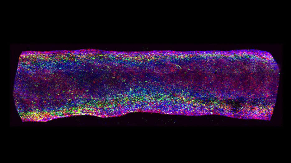
Microscopic image of a segment of bile duct in Dr. Ogawa’s model reveals cell cilia (red and green). DNA (blue) is visible in all cells. This new model may support the identification of more effective therapies and improve understanding of disease progression, with the potential to inform strategies that prevent advanced disease. (Image c/o Ogawa Lab)
A new lab-grown model of bile ducts inside the liver could help researchers better understand how biliary diseases develop and progress.
The biliary system—the part of the digestive system that includes the gallbladder, bile ducts, and parts of the liver—relies on a complex set of chemical and physical signals to function. Recreating this complexity in the lab has been a long-standing challenge, limiting researchers’ ability to understand how the system works and how diseases arise.
Now, Dr. Shinichiro Ogawa and a team at UHN’s McEwen Stem Cell Institute (McEwen) have developed a model that more closely reflects how the biliary system works than ever before. This advancement could help researchers to better study and eventually treat biliary diseases.
“To date, even advanced models of the bile ducts are not comprehensive,” says Dr. Ogawa, the study’s senior author. “Some models show how bile flow affects cell functioning. Others are better for studying what’s happening inside the cells. Our goal was to create a new 3D system that integrates all these features and allows us to better understand how cholangiocytes—bile duct cells—develop, function, and become diseased.”
To do this, the researchers used stem cells and a specialized system called an AngioPlate to generate three-dimensional bile ducts in the lab. They then assessed how well these structures functioned and responded to stress.
The new model recapitulated features of both healthy and diseased bile duct physiology. It included functional hallmarks such as cilia—hair-like structures that help move fluid through the ducts—and a network of supportive stromal cells that contribute to duct structure and function.
When exposed to disease-associated factors, such as bile acids and inflammatory molecules called cytokines, the model exhibited changes consistent with biliary diseases. These included impaired barrier integrity and bile leakage—key features of disease progression. Importantly, the platform also enabled the McEwen team to observe how disease unfolds over time, capturing multiple stages of its development.
By more accurately reflecting the complexity of the bile duct environment, this model offers a more realistic platform to study both healthy bile duct function and disease progression. It offers researchers a way to better understand how these tissues develop, respond to stress and injury, and fail. This new approach also lays the groundwork to enable patient-specific models, bringing us closer to more targeted treatments and improved outcomes for people with biliary diseases.
Britney Tian, a graduate student researcher at McEwen Stem Cell Institute, is the first author.
Dr. Shinichiro Ogawa is the senior author on this publication. He is a Scientist at UHN’s McEwen Stem Cell Institute and Assistant Professor at the University of Toronto’s Temerty Faculty of Medicine in the Department of Laboratory Medicine & Pathobiology.
This work was supported by the University of Toronto Medicine by Design initiative, the Canada First Research Excellence Fund, the Canadian Institutes of Health Research, JSPS KAKENHI, the Stem Cell Network, and UHN Foundation.
For a list of competing interests, please see the publication.
-
Tian B, Ogawa M, Kondo M, Langeveld G, Huan LJ, Zhang F, Hollinger A, Deir S, Bear C, Zhang B, Ogawa S. Bioengineered 3D hPSC-Cholangiocyte Ducts With Physiological Signals for Biliary Disease Modeling. Adv Healthc Mater. 2026 Mar 18:e05293. doi: 10.1002/adhm.202505293.