Brain Plasticity and Chronic Pain
Researchers identify brain changes associated with pain relief after TN treatment.
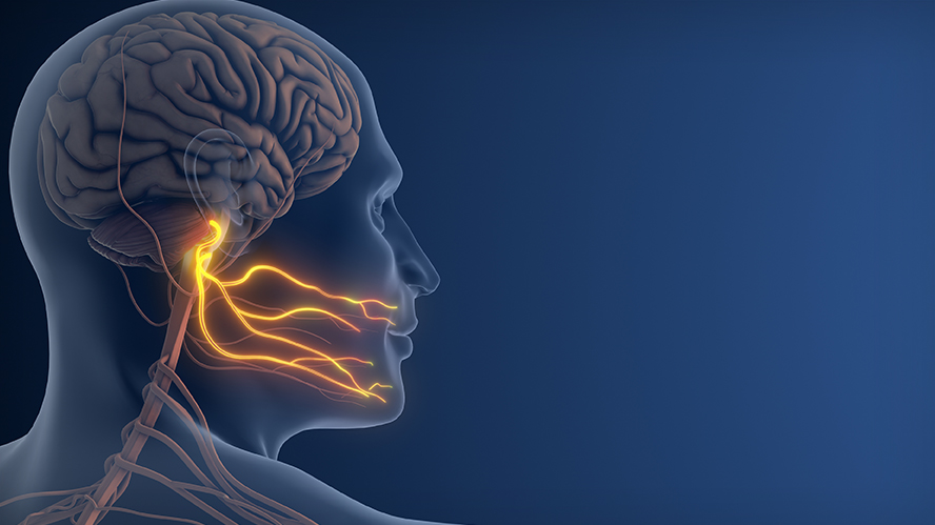
Up to 25% of the world is affected by chronic pain conditions such as trigeminal neuralgia (TN)—a form of chronic nerve pain affecting the face and mouth. For those who fail to get relief from standard treatments, understanding why is an essential first step to improving care. (Image: Getty)
A new study from a team at UHN's Krembil Brain Institute (KBI), led by Senior Scientist Dr. Mojgan Hodaie, is shedding light on how specific structural changes in the brain, a form of brain (or neuro) plasticity, are associated with pain relief in trigeminal neuralgia (TN) following surgical treatment. For individuals living with TN—severe chronic neuropathic (nerve) facial pain—these findings may help advance personalized treatment planning and improve understanding of the brain's response to pain relief.
For many patients, trigeminal neuralgia (TN) can be effectively managed with surgery. However, about 25% of individuals do not respond well to surgery. Understanding why remains a challenge in helping more patients achieve effective pain relief.
Prior research from Hodaie's team revealed changes in the anatomy of 13 brain regions among people with TN that could help predict whether a patient was likely to respond to surgery. These regions showed reduced grey matter volume and reduced thickness of the cortex—the outermost layer of the brain. "We wanted to better understand what happens to these regions after treatment, and whether there is a correlation with pain relief," says Dr. Hodaie.
To explore the connection between brain changes and TN symptoms, KBI researchers evaluated MRI scans and pain relief scores from 115 patients with TN before and after surgical treatment.
Comparing imaging before and after surgery, the team observed changes in several brain structures. They identified eight key brain regions where increased thickness of the cortex after surgery was related to improvements in TN symptoms. In general, they found that in these regions, greater thickening between pre- and post-surgery was associated with greater pain relief. One region, the fusiform gyrus—an area of the brain linked to pain-memory—showed the strongest correlation, with post-surgical thickening of the cortex directly linked to pain relief achieved.
These findings give health care providers new insights into the biological processes underlying pain relief in TN. Moreover, they offer evidence that the brain's ability to change and adapt in response to experiences and conditions, a process known as neuroplasticity, may play a key role in the brain's response to chronic pain. For people with TN, this work lays the groundwork for the development of more personalized approaches to treatment in the future. Additional studies will further investigate the role of the fusiform gyrus and explore pain relief from a more multifaceted lens.
Emili Adhamidhis is the first author of this study. She is a medical student at McMaster University's Michael G. DeGroote School of Medicine and a former research trainee who obtained her MSc at UHN's Krembil Brain Institute.
Dr. Mojgan Hodaie, the senior author of this study, is a Senior Scientist at UHN's Krembil Brain Institute, Affiliate Scientist at UHN's KITE Research Institute, and Professor of Surgery (Neurosurgery) at the University of Toronto's Temerty Faculty of Medicine.
This work was supported by the Institute of Medical Science and Temerty Faculty of Medicine at the University of Toronto, and UHN Foundation.
Adhamidhis E, Srisaikaew P, Li J, Latypov TH, Noorani A, Hodaie M. Brain Regional Predictors of Surgical Response Are Dynamically Associated with Post-Surgical Pain Relief in Trigeminal Neuralgia. Brain Sci. 2026 Sep 13;16(9):966.