

A new Toronto General Hospital Research Institute (TGHRI) study has shed light on how cells in blood vessels communicate with each other, paving the way for new treatments for cardiovascular diseases like atherosclerosis—the build-up of fats, cholesterol, and other substances on the artery wall. Atherosclerosis can cause arteries to narrow, blocking blood flow and potentially lead to conditions such as heart attack or stroke.
Endothelial cells (ECs) line the inner surface of blood vessels, including arteries. Dysfunctional endothelium—changes to EC structure and function—is often the earliest disruption in numerous cardiovascular diseases. ECs communicate by releasing small packages called extracellular vesicles (EVs), which carry essential molecules like proteins capable of influencing the biological activities of recipient cells.
“We know that ECs release vesicles that contain different components when they are activated versus inactivated. EC activation can happen due to stimuli such as inflammation in conditions like atherosclerosis. Despite ECs residing between the bloodstream and vessel wall, it remains uncertain whether they release vesicles in a directional manner to convey specific messages to these parts,” says Dr. Kathryn Howe, Scientist at TGHRI, Vascular Surgeon at the Peter Munk Cardiac Centre, and senior author of the study.
To determine this, the researchers isolated EVs from human aortic endothelial cells and analyzed their contents. They found that when these cells were activated, as occurs in conditions like atherosclerosis, they released more EVs containing molecules linked to the disease such as regulators of inflammation.
These vesicles talk to immune cells (monocytes) and muscle cells found in the blood and vessel walls, causing changes to their genetic instructions which then impact cell behaviour.
“Importantly, we discovered that endothelial cells predominantly release more vesicles from the side facing the bloodstream, known as the apical side, compared to the side facing the vessel wall, known as the basolateral side. This pattern increased with activation,” says Dr. Sneha Raju, first author of the study.
“The vesicles that were delivered from the apical side had different cargo than the ones coming from the basolateral side,” adds Dr. Raju who is a PhD Candidate co-supervised by Dr. Howe and Dr. Jason Fish, Senior Scientist at TGHRI and co-author of this study.
Further analysis demonstrated that upon activation, both apical and basolateral messages undergo alterations, with those directed towards the vessel wall exhibiting the most pronounced changes associated with the development of artery disease. This activated messaging changes gene expression in neighbouring cells.
This discovery helps us better understand how cells communicate and opens up new possibilities for the design of endothelial-based therapeutics for cardiovascular diseases such as atherosclerosis where ECs are persistently activated.
This work was supported by the Canadian Institutes of Health Research, National Institutes of Health, Heart and Stroke Foundation of Canada, Vascular Cures, Bill and Vicky Blair Foundation, Peter Munk Cardiac Centre, the Canada Foundation for Innovation, Natural Sciences and Engineering Research Council of Canada, the Canada Research Chairs program, and UHN Foundation. Dr. Kathryn Howe is an Associate Professor in the Department of Surgery at the University of Toronto.
Co-author of the study, Dr. Natalie Galant is a cofounder and CEO of Paradox Immunotherapeutics.
Raju S, Botts SR, Blaser MC, Abdul-Samad M, Prajapati K, Khosraviani N, Ho TWW, Breda LCD, Ching C, Galant NJ, Fiddes L, Wu R, Clift CL, Pham T, Lee WL, Singh SA, Aikawa E, Fish JE, Howe KL. Directional Endothelial Communication by Polarized Extracellular Vesicle Release. Circ Res. 2024 Feb 2;134(3):269-289. doi: 10.1161/CIRCRESAHA.123.322993.
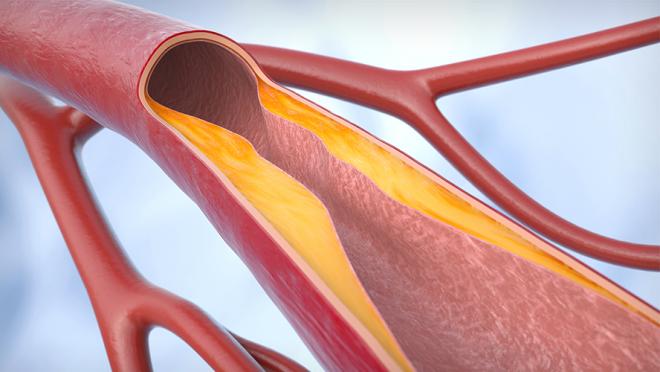
In atherosclerosis, there is a gradual buildup of plaque (fats, cholesterol and other substances) inside the walls of arteries due to inflammation. This reduces blood flow to the vital body organs. (Getty Images)