


Image-guided cancer therapies rely on the ability to visualize a malignant tumour so that treatment is applied only at that location. This is achieved by injecting a "contrast agent"—a compound that selectively accumulates in cancerous tissue and can be observed by medical imaging methods.
One of the most highly customizable types of contrast agents is a microbubble. These bubbles are very small (1 to 5 microns in diameter) and consist of a shell and gas core. Although they can be readily detected by ultrasound imaging, microbubbles burst when they are exposed to ultrasound. This makes them unusable for imaging purposes, as they can only help visualize tumours over a short window of time.
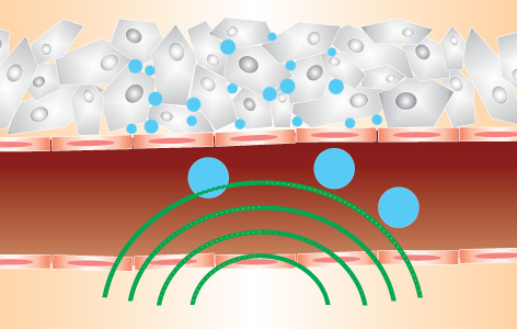
To address this issue, Techna Core Lead and PM Senior Scientist Dr. Gang Zheng invented microbubbles made of a compound called porphyrin. Porphyrin microbubbles still burst when they are exposed to ultrasound imaging, but their remnants form porphyrin nanoparticles that stay within the tumour and can be visualized. Microbubbles are versatile; they can be loaded with drugs within the gas core and when they burst, the drug is delivered directly to the tumour. Moreover, they can be visualized using imaging methods other than ultrasound.
"To our knowledge, this is the first study to explore the conversion of microbubbles into nanoparticles," says Dr. Zheng. "These improved microbubbles can be visualized for longer, enabling clinicians to apply treatments more accurately and minimize damage to the surrounding healthy tissue. Coupled with the fact that microbubbles can act as drug carriers, they represent a powerful new tool with which to specifically target cancerous tumours."
This work was supported by the Canadian Institutes of Health Research, the Canadian Space Agency, the Natural Sciences and Engineering Research Council of Canada, the Ontario Institute for Cancer Research, the Ministry of Knowledge Economy, South Korea, the Joey and Toby Tanenbaum/Brazilian Ball Chair in Prostate Cancer Research, the Canada Foundation for Innovation and the Princess Margaret Cancer Foundation.
In situ conversion of porphyrin microbubbles to nanoparticles for multimodality imaging. Huynh E, Leung BY, Helfield BL, Shakiba M, Gandier JA, Jin CS, Master ER, Wilson BC, Goertz DE, Zheng G. Nature Nanotechnology. 2015 Mar 30. [Pubmed abstract]