

Home page Description:
Researchers find brain regions implicated in the development of fatigue in Parkinson disease.

Posted On: November 30, 2016
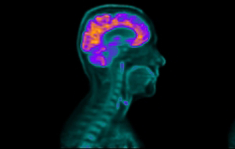
Image Caption:
Brain regions that are more active during a positron emission tomography (PET) scan show up as brighter in the resulting brain image (pictured above).
Parkinson disease (PD) is the second most common neurodegenerative disorder after Alzheimer disease. It is characterized by tremors, rigid muscles, slow movements and problems with posture and balance. In addition to these symptoms, up to 70% of people living with PD will experience an unexplainable and debilitating fatigue, for which no treatment is available.
Krembil Senior Scientist Dr. Antonio Strafella launched a study to find out why so many people with PD feel an overwhelming lack of energy. He used a positron emission tomography scanner to obtain brain images from 23 PD patients with and without fatigue. Positron emission tomography is an advanced imaging technique that can be used to examine the metabolic activity (ie, energy use) of tissues comprising an organ such as the brain. Study participants also completed questionnaires to assess their fatigue level and sleep quality.
By comparing the brain images, the research team discovered that two groups of brain regions in particular—the salience network and the default mode network—display different levels of activity between PD patients with and without fatigue. The salience network was less active and the default mode network was more active in PD patients with fatigue than in those without fatigue. The salience network is involved in preparing the brain for action, so its reduced activity may be a contributing factor in causing fatigue in PD.
“According to the patients in my clinical practice, fatigue is one of the more devastating symptoms of PD,” says Dr. Strafella. “However, we must work to better understand the physiological mechanisms that contribute to fatigue in PD before we can develop a way to treat it.”
This work was supported by Parkinson Society Canada, and the Toronto General & Western Hospital Foundation. AP Strafella holds a Tier 2 Canada Research Chair in Movement Disorders and Neuroimaging.
Fatigue in Parkinson's disease: The contribution of cerebral metabolic changes. Cho SS, Aminian K, Li C, Lang AE, Houle S, Strafella AP. Human Brain Mapping. doi: 10.1002/hbm.23360. 2016 August 29. [PubMed abstract]