


A recent study conducted at UHN’s Donald K. Johnson Eye Institute (DKJEI), in collaboration with The Hospital for Sick Children (SickKids), has provided valuable insights into why individuals with Roifman syndrome experience vision-related symptoms.
Roifman syndrome is a rare genetic disorder characterized by abnormal growth of the bones and joints, disrupted immune function and cognitive delay. Individuals with the condition also have vision problems caused by degeneration of the retina—the thin layer of light-sensing tissue that lines the back of the eye.
Due to the rarity of this condition, very little is known about the features of associated eye disease.
“Our limited understanding of retinal degeneration in this condition hinders our ability to diagnose patients and manage their symptoms,” explains Dr. Brian Ballios, a DKJEI Scientist and the lead author of the study. “We need a deeper understanding of underlying eye disease processes to better help our patients.”
To address this knowledge gap, the team conducted a detailed structural and functional evaluation of the retina in ten people with Roifman syndrome. The team used multiple research techniques, including electroretinography to measure the electrical activity of retinal cells, and fundus autofluorescence imaging to visualize the structure and health of retinal cells.
“We discovered that the most common structural abnormality was a ring of heightened autofluorescence surrounding the central part of the retina,” says Dr. Ajoy Vincent, a Staff Ophthalmologist at SickKids and the senior author of the study. “This ring indicates abnormalities in the retinal pigment epithelium—a layer of cells that support the function and health of specialized neurons called photoreceptors. These photoreceptors are essential for vision and convert light into electrical signals.”
In line with these findings, many individuals with Roifman syndrome have photoreceptor dysfunction and corresponding visual impairments such as night blindness.
Follow-up studies conducted over several years revealed that some patients experience progressive retinal atrophy and decreased visual acuity, suggesting that the retinal degeneration associated with Roifman syndrome worsens slowly over time.
The team also found that age alone is not a key determinant of disease severity, highlighting the need to study other factors that might contribute to symptoms, such as sex and specific mutations in the Roifman gene.
This study lays a strong foundation for future research into the underlying mechanisms of Roifman syndrome and for the development of targeted treatments.
“It was immensely gratifying to conduct this research in collaboration with Dr. Chaim Roifman. Dr. Roifman is a world-leading paediatric immunologist, and he was the first to describe the clinical features of the disorder in 1999,” says Dr. Ballios. “His expertise helped us understand how the specific features of eye disease fit into the bigger clinical picture of this disorder by considering patients’ overall health concerns.”
This work was supported by the Foundation Fighting Blindness U.S. and the UHN Foundation. Drs. Ballios and Vincent are Assistant Professors in the Department of Ophthalmology and Vision Sciences at the University of Toronto.
Ballios, B. G., Mandola, A., Tayyib, A., Tumber, A., Garkaby, J., Vong, L., Heon, E., Roifman, C. M., & Vincent, A. (2023). Deep phenotypic characterization of the retinal dystrophy in patients with RNU4ATAC-associated Roifman syndrome. Eye (London, England), 2023 May 24. doi: 10.1038/s41433-023-02581-1.
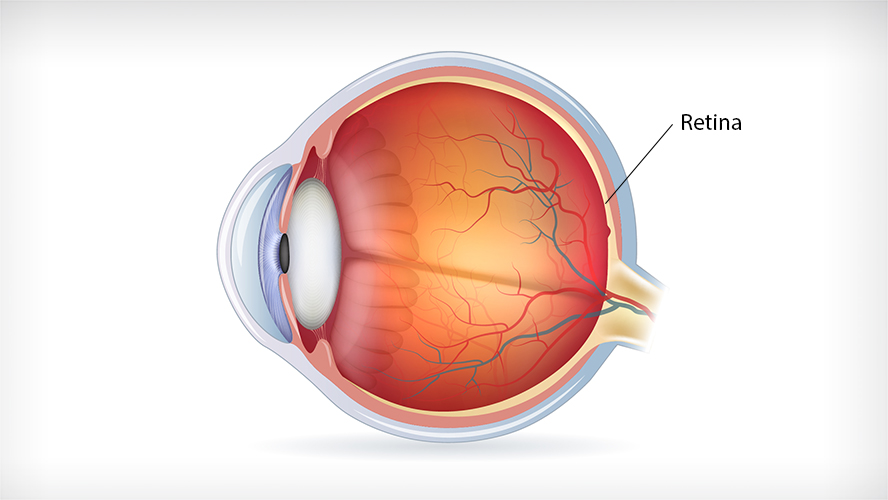
Photoreceptors are specialized neurons that detect light and convert it into electrical signals that are transmitted to the brain. These cells are found in the retina, the thin layer of tissue that lines the back of the eye.