


Researchers at the University of Toronto and University Health Network have uncovered new information that could accelerate post-surgical healing for procedures involving medical devices as diverse as dental implants and skin dressings.
Their paper, published in Nature Communications Biology describes how different surface textures on medical implants affect a fundamental process in the body’s ability to heal the area surrounding the implant.
“We have known for decades that creating nano-scale implant surface texture improves clinical success rates,” said John E. Davies, a professor in University of Toronto’s Faculty of Dentistry and Institute of Biomaterials & Biomedical Engineering (IBBME)—the senior author of this study. “However, little was known of the cellular mechanisms by which the implant surface affects the healing process.”
The team was co-led by Dr. Ralph DaCosta, a Scientist at Princess Margaret Cancer Centre and Research Faculty at the Techna Institute at University Health Network, who looked at the effect of implant surface texture on neovascularization—the process of blood vessel formation— around implants.
“The ability for blood vessels to form around an implant is a key factor in its successful tissue integration,” said Niloufar Khosravi, a PhD candidate in Davies’ lab and the study’s first author. “Indeed, neovascularization is vital for tissue healing and regeneration around the implant.”
To study this effect, the team built a model that allowed them to observe the implant healing process in real-time using intravital optical microscopic imaging systems developed in DaCosta’s lab. Their experiments monitored the formation of new blood vessels around a series of medical-grade titanium implants at the cellular level, and how the distribution of these vessels changed according to different surface textures on the implants.
“We did not initially expect blood vessels to be clearly visible in this model,” said Khosravi. “We decided to add an imaging contrast agent after a few pilot studies and the resulting resolution was remarkably higher than we expected. It allowed us to monitor not only the rate, but also the location, shape, function and pattern of new blood vessels around implants over a six week period.”
In addition to providing new insights into how medical implants can be better designed, the team’s findings could also address other health related challenges.
“We can use this knowledge to build broader implantable biomaterials to improve the quality of life for people who have impaired healing conditions, such as the elderly or persons with diabetes,” said Dr. DaCosta, who is also an assistant professor in the University of Toronto’s Department of Medical Biophysics. “We now have new tools and strategies to better support the body’s natural repair process.”
Story source: Luke Ng, University of Toronto.
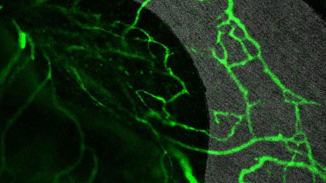
Blood vessels (green) forming on a nano-surfaced titanium implant (grey) as visualized by intravital microscopy using the team’s model. (Image: Niloufar Khosravi).