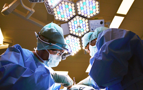
Advanced imaging techniques have been developed that give surgeons more precise control when removing tumours from locations that are complex and hard to identify.
For example, prior to cancer surgery, x-ray computed tomography (CT) is often used to construct a detailed three-dimensional image to help the surgeon visualize the tumour within complex healthy structures and to plan the optimal surgical procedure. Another imaging technique called fluorescence optical imaging is also being increasingly used for identification and surgical removal of cancer that has spread to the lymph nodes.
Until now, the combined use of these techniques in the operating room for tumour and lymph node visualization has been limited because multiple imaging agents are needed and existing agents do not have sufficient sensitivity and specificity for disease detection. Techna Scientist Dr. Jinzi Zheng and her collaborators may have found a way to overcome this limitation by developing a single injectable imaging agent that can be used for CT and fluorescence imaging of tumours and malignant lymph nodes.
Dr. Zheng and her team developed the agent by encapsulating, into a single nanoparticle, different imaging molecules that were engineered to meet the clinical requirements for CT and fluorescence imaging. Data obtained from various experimental cancer models showed that the new imaging agent improved sensitivity when mapping the location of the tumour.
Explains Dr. Zheng, "This new technology is particularly useful because it enables us to employ different imaging techniques prior to and during surgery following one injection of the imaging agent. Our preliminary results are particularly promising and suggest that this agent could be used to improve the localization, detection and removal of a wide range of cancers."
This work was supported by the Fidani Family Chair in Radiation Physics, the Kevin & Sandra Sullivan Chair in Surgical Oncology, the RACH Fund and The Princess Margaret Cancer Foundation.
A multimodal nano agent for image-guided cancer surgery. Zheng J, Muhanna N, De Souza R, Wada H, Chan H, Akens MK, Anayama T, Yasufuku K, Serra S, Irish J, Allen C, Jaffray D. Biomaterials. 2015 Jul 14. [Pubmed abstract]


