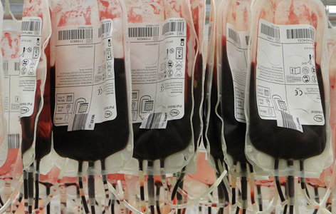
Thalassemia major is a hereditary condition where people lack the ability to produce normal hemoglobin—the oxygen-carrying molecule in blood—causing chronic fatigue. It requires regular blood transfusions to treat. A common side effect of these transfusions is an overload of iron in the body, which can damage the heart and other organs. Magnetic resonance imaging (MRI) is being used clinically to monitor for potential iron overload with a specific focus on the liver and the heart; finding such an overload may trigger initiation of or changes in chelation therapy.
A recent study by Techna Affiliated Faculty Dr. Bernd Wintersperger and colleagues used magnetic resonance imaging (MRI) to further examine the hearts of patients with thalassemia major in detail. MRI enabled the researchers to take readings for what is known as the extracellular volume (ECV) fraction, a measure that reflects the amount of space outside the muscle cells in the heart. In various cardiac pathologies ECV has been shown to reflect the degree of subtle diffuse scaring of the heart muscle; such changes are associated with further damage of the muscle resulting in impaired cardiac function and potentially life-threatening heart rhythm irregularities. In this study, patients with a history of iron overload had a higher ECV measurement—indicative of early damage—while patients without prior iron overload or healthy individuals did not.
Heart failure is one of the major causes of death in patients with iron overload, and once damage to the heart begins it cannot be reversed. Early detection of damage with MRI will allow medical care teams to better manage iron levels in patients with thalassemia major and other conditions that require regular blood transfusions to help prevent damage to the heart. These findings suggest that MRI could serve as an effective way to monitor for the earliest signs of damage.
"We clinically apply MRI techniques that let us measure iron overloads in the heart, but those overloads may be reversible by therapeutic interventions. However, this technique provides us further non-invasive insight into resulting damage of the heart muscle which may currently may not be reversible. This may potentially result in earlier triggers for intervention," comments Dr. Wintersperger.
This work was supported by the Radiological Society of North America, the Toronto General & Western Hospital Foundation and by non-financial research support from Siemens.
Quantification of myocardial extracellular volume fraction with cardiac MR imaging in thalassemia major. Hanneman K, Nguyen ET, Thavendiranathan P, Ward R, Greiser A, Jolly MP, Butany J, Yang IY, Sussman MS, Wintersperger BJ. Radiology. 2015 Dec 10. [Pubmed abstract].


