

Home page Description:
Technologies combined to rapidly identify tumours that will need more aggressive treatment.

Posted On: March 06, 2017
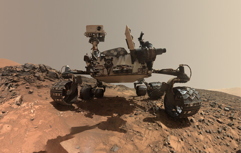
Image Caption:
Mass spectrometry, which has potential health care applications, was also used by the Mars Curiosity rover (pictured) to look for the chemical fingerprints of life.
The best strategy to treat cancer for a particular patient depends on many factors, including the characteristics of the tumour. Tumours that contain necrotic tissue (ie, cells killed by environmental factors, such as a lack of oxygen) require more aggressive treatment. When surgeons remove a tumour from a patient, the tissue is tested by pathologists to check for necrosis using a microscope. That process can take up to half an hour—time that the patient spends under anesthesia in an operating room.
Techna’s Dr. Arash Zarrine-Afsar and colleagues believe that a technology called mass spectrometry can be used to more rapidly check for necrosis, and to distinguish cancer cells from healthy tissue. Mass spectrometry involves extracting a sample of tissue, then analyzing the chemical components to produce a “chemical fingerprint” that is unique to each type of tissue (necrotic, healthy, cancerous, etc.). The challenge in applying the technique clinically is that mass spectrometry is no faster than traditional microscope-based pathology when the entire surface of the tissue removed during surgery has to be tested.
Adding another technique based on polarized light imaging (polarimetry) can overcome this hurdle. Polarimetry can instantly identify tumour regions with suspected necrosis. These regions can then be rapidly tested in a more focused and accurate way using mass spectrometry. Using an experimental model, the Techna scientists confirmed that the combined method could identify necrotic tissue in minutes.
Use of this technique may help surgical teams to more rapidly adjust treatment plans in response to the presence of necrosis in a tumour. It may also help to cut health care costs and increase patient convenience by reducing the amount of time that patients spend in the operating room.
This work was supported by The Princess Margaret Cancer Foundation.
Tata A, Woolman M, Ventura M, Bernards N, Ganguly M, Gribble A, Shrestha B, Bluemke E, Ginsberg HJ, Vitkin A, Zheng J, Zarrine-Afsar A. Rapid Detection of Necrosis in Breast Cancer with Desorption Electrospray Ionization Mass Spectrometry. Sci Rep. 2016 Oct 13. doi: 10.1038/srep35374.