


Posted On: January 02, 2015
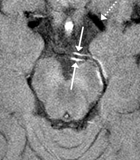
Image Caption:
VW-MRI image showing a normal blood vessel which is black (dotted arrow) compared with an abnormal blood vessel which has brightness in the vessel wall indicating inflammation (solid arrows).
Vessel wall magnetic resonance imaging (VW-MRI) is a medical imaging technique being developed to more accurately diagnose an ischemic stroke—a stroke caused by a blocked blood vessel in the brain. Traditionally, physicians have determined the cause of a stroke using images of the blood flowing inside of a blood vessel. However, many of the diseases that cause stroke alter the wall of the vessel and not the blood per se. VW-MRI generates detailed three-dimensional images of the wall of a blood vessel, enabling radiologists to see more clearly what the underlying disease is: a blood clot, fatty plaques, or inflammation or constriction of the blood vessel. Ischemic stroke can be treated with different medications and/or surgery, depending on its cause.
TWH neuroradiologist Dr. Daniel Mandell is developing VW-MRI and focusing on improving the clinical interpretation of its images. Recently, he discovered that the minimally invasive surgical removal of a blood clot (to treat ischemic stroke) can cause changes in the blood vessel wall which mimic other artery diseases.
As part of his clinical study, 16 patients who had suffered an ischemic stroke underwent VW-MRI within days of being treated. The imaging showed a thickening and brightening of the blood vessel wall more frequently in stroke patients treated with surgery than in those treated with medications alone. These same features are also observed on VW-MRI images of blood vessels damaged by inflammatory diseases.
The findings may help to prevent the misdiagnosis of artery disease in people who have been treated for ischemic stroke. Furthermore, the accurate interpretation of VW-MRI images will help clinicians to make better diagnoses of ischemic stroke and prescribe the most effective therapies to treat it.
This work was supported by The Association of University Radiologists and the Toronto General & Western Hospital Foundation.
Vessel wall magnetic resonance imaging in acute ischemic stroke: effects of embolism and mechanical thrombectomy on the arterial wall. Power S, Matouk C, Casaubon LK, Silver FL, Krings T, Mikulis DJ, Mandell DM. Stroke. 2014 August [Pubmed Abstract]
Comments