


By: Dr. Laura Kuhlmann, ORT Times Science Writer
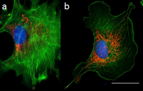
In fluorescence microscopy, fluorophores (chemical compounds which re-emit light upon excitation by a light source such as a laser) are used to label structures/molecules of interest and achieve contrast in biological samples. In thicker specimens, however, out-of-focus light can create hazy images and decrease contrast. These problems are mitigated in confocal microscopy, which uses a pinhole to block out-of-focus light, thus increasing the resolution of thick specimens.
Confocal microscopy is among the most requested services at the Advanced Optical Microscopy Facility (AOMF) at UHN. They offer users a diverse range of confocal microscopes to choose from such as confocal laser-scanning microscopes (CLSM) and stimulated emission depletion (STED) microscopes, which offer higher resolution (i.e. super-resolution). However, confocal microscopy handbooks are often overwhelming for biologists and many users encounter difficulties when troubleshooting their experiments, which can compromise image quality and data quantification.
Responding to a need for comprehensive tutorials, James Jonkman, manager of AOMF, and collaborators put together a Nature Protocols paper offering step-by-step guidance for quantitative confocal microscopy. The article covers how confocal microscopes work, how to choose the most appropriate instrument for your experiment and how to prevent bias. The article also describes sample preparation hazards and includes key references for users wanting to follow up on these topics.
Preventing bias in confocal microscopy when selecting an area to image and quantify is not trivial. “Systematic ways of choosing the cells or sampling are really important but are often overlooked,” Jonkman says. There is a high risk that “your eyes are drawn to the fields of view that match your hypothesis.” Jonkman offers impartial data acquisition strategies for confocal microscopy in the new publication.
Another important aspect is optimizing your imaging parameters as discussed in Intro to AOMF – Part 1. “It is important to not treat the CLSM like a routine scanner.” Ask the AOMF staff for advice when varying acquisition parameters.
In addition, the paper addresses instrumentation problems—which could jeopardize image quality—that most users are not aware of. Examples include focus drift with the STED microscope and laser intensity fluctuations in CLSM. While these problems can be mitigated by implanting longer instrument warm-up times, a solution like this may not be feasible at a busy core facility. Combined with inherit variability in biological samples, these fluctuations further complicate data quantification. “We need to do a better job of teaching the users about these issues,” Jonkman says. “We need to convince the companies to change the instruments.” The authors recommend that manufacturers incorporate monitoring devices (e.g., laser power meters) into microscopes to enable hardware and software corrections in real time. These changes could improve rigour and reproducibility in confocal microscopy while making the imaging systems more user-friendly.