 NRx (formerly known as Net Results Express) is UHN's monthly research e-newsletter. Through NRx you can read about ongoing research at our five research institutes, the Ontario Cancer Institute (OCI), the Toronto General Research Institute (TGRI), the Toronto Western Research Institute (TWRI), the Toronto Rehabilitation Institute (TRI) and the Techna Institute (Techna).
In this issue you can read about research in:
- The regulation of blood cell development
- Preventing the rejection of cardiac stem cell transplants
- Mind wandering and the perception of pain
- Measuring brain waves
- Reducing the production of glucose in the liver
- Protection of cognitive function after brain injury
We hope that you will find NRx informative. If you have feedback or questions, please contact www@uhnresearch.ca.
Christopher J. Paige, PhD, FCAHS
Vice President, Research
University Health Network
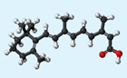
In addition to blood cell development, retinoic acid (depicted above) plays a role in the formation of the central nervous system.
A recent study by McEwen Centre for Regenerative Medicine Director and OCI Senior Scientist Dr. Gordon Keller has brought new insight into how blood-forming stem cells are generated by identifying a key factor that regulates their development in the early embryo.
Using various experimental models, Dr. Keller and his team demonstrated that the molecule retinoic acid (RA) enhances the development of blood-forming stem cells. The team also discovered that inactivating the protein responsible for production of RA in a population of specialized stem cell inhibited the development of blood-forming stem cells.
Dr. Keller explains, "Our findings have identified a critical regulator for directing pluripotent stem cells to make blood-forming stem cells. This brings us one step closer to our goal of developing a new and unlimited source of these stem cells for transplantation for the treatment of different blood cell diseases."
This work was supported by the Canadian Institutes of Health Research and the National Institutes of Health.G. Keller is a Tier I Canada Research Chair in Embryonic Stem Cell Biology.
Retinoic acid signaling is essential for embryonic hematopoietic stem cell development. Chanda B, Ditadi A, Iscove NN, Keller G Cell. 2013 September 26. [Pubmed abstract]

Heart attacks cause irreparable damage to cardiac tissue leading to the formation of scar tissue that does not function normally.
Mesenchymal stem cells (MSCs) have been found to improve heart function when they are transplanted into patients with injured hearts. However, MSCs from older patients have limited capacity for tissue regeneration and restoration of heart function. Stem cells from young donors have a greater regenerative capacity and may be better candidates for clinical application. Unfortunately, these allogeneic stem cells—referred to as allogeneic because they come from a genetically non-identical donor—can only evade the immune system early after implantation and are eventually killed by the recipient’s immune cells.
In an attempt to improve the survival of transplanted MSCs and increase their potential to repair damaged hearts, TGRI Senior Scientist Dr. Ren-Ke Li and his collaborators have examined which factors might help MSCs maintain their ability to not activate the recipient’s immune system. Dr. Li and his team found that a protein known as prostaglandin E2 (PGE2) is able to induce the expression of proteins that suppress immune responses. They were was also able to show that when PGE2 is slowly released at the site of MSC injection, through the use of a biodegradable gel, the transplanted stem cells survive longer and improve heart function.
Explains Dr. Li, "We demonstrated that maintaining PGE2 levels prevents the rejection of MSCs and leads to improved heart function. Hopefully, these findings will be useful in developing allogeneic stem cell-based cardiac therapies that can improve the current standard of care for cardiovascular diseases."
This work was supported by the Canadian Institutes of Health Research. RK. Li is a Tier I Canada Research Chair in Cardiac Regeneration.
Preserving prostaglandin e2 level prevents rejection of implanted allogeneic mesenchymal stem cells and restores postinfarction ventricular function. Dhingra S, Li P, Huang XP, Guo J, Wu J, Mihic A, Li SH, Zang WF, Shen D, Weisel RD, Singal PK, Li RK. Circulation. 2013 September 10. [Pubmed abstract]

Humans spend half their time on thoughts not associated with their immediate sensory environment, like daydreaming, planning and revisiting memories.
In day-to-day situations, the human mind often wanders. But it is always assumed that painful sensations automatically capture attention. TWRI Senior Scientist Dr. Karen Davis and her doctoral student Aaron Kucyi recently published the results of a study examining how the mind wanders when perceiving pain.
The study demonstrated that there is a wide variety in perception of pain between individuals, with some people always paying attention to pain while others who often drift away in thought. These differences in attention were found to be linked with the function and structure of the pain- and attention-related regions of the brain.
When people’s minds wander away from pain, there is increased activation in the default mode network (DMN), regions of the brain implicated in mind wandering, and strong interactions between the DMN and periaqueductal gray (PAG), an endorphin-containing region of the brain that suppresses pain. Individuals with greater tendencies to mind wander from pain had stronger anatomical links and more flexible communication between PAG and DMN.
Commenting on the study, Dr. Davis says, "These findings provide clinically important clues about why some individuals cannot disengage from pain, such as patients suffering from chronic pain. However, the findings also suggest that the brain circuitry in other individuals with pain may allow them to control their pain through a variety of therapeutic strategies, including cognitive-behavioural therapy."
This work was supported by the Canadian Institutes of Health Research.
Mind wandering away from pain dynamically engages antinociceptive and default mode brain networks. Kucyi A, Salomons TV and Davis KD. Proceedings of the National Academy of Sciences. 2013 October 28. [Abstract]

Changes in neural oscillations can accompany a number of neurological diseases including epilepsy, schizophrenia and Parkinson’s disease.
Measurable electrical neural oscillations are signals that represent the activity of the central nervous system. They communicate information between regions of the brain. Currently, the best methods of studying the brain circuitry responsible for generating neural oscillations come from animal models—no equivalent human models exist.
TWRI Affiliate Scientist Dr. Taufik Valiante, in collaboration with TWRI Affiliate Scientist Dr. Liang Zhang and TWRI Senior Scientist Dr. Peter Carlen, has demonstrated a new technique that can maintain human brain samples outside the body—these sections of brain tissue can generate neural oscillations similar to those observed in the living brain. These samples, known as neocortical slices, are obtained from a surgical procedure that removes tissue from the neocortex of patients with epilepsy.
Two types of neural oscillations were observed in the neocortical slices—theta oscillations and high gamma waves—which are thought to be involved in a number of cognitive and motor functions. These oscillations are difficult to generate in existing animal models, and thus have not been extensively studied in neocortical slices. Dr. Valiante says, "We observed that these two waves were coupled together, and to one another, suggesting a novel type of communication between layers of the neocortex. This technique will allow us to better investigate the circuitry of the human brain."
This work was supported by the Canadian Institutes of Health Research.
In vitro recordings of human neocortical oscillations. Florez CM, McGinn RJ, Lukankin V, Marwa I, Sugumar S, Dian J, Hazrati LN, Carlen PL, Zhang L, Valiante TA. Cerebral Cortex. 2013 September 17. [Pubmed abstract]

Obesity is a major risk factor for the development of type 2 diabetes.
Glucagon-like peptide-1 (GLP-1) is a gastrointestinal hormone that has important implications in controlling blood glucose (sugar) levels. In addition to stimulating insulin secretion upon food intake, GLP-1 is associated with many other beneficial effects—including inducing the feeling of fullness after eating, reducing gastric emptying, regulating metabolism and improving cardiac and vascular function—such that anti-diabetic drugs targeted at increasing its activity have been developed. A recent study from TGRI Scientist Dr. Tianru Jin and his team shows that GLP-1(28-36)a, a "by-product" of GLP-1, also has beneficial effects on repressing glucose production in the liver.
Using an experimental model of diet-induced obesity, the team showed that administration of GLP-1(28-36)a resulted in a significant reduction in body weight gain compared with controls. Glucose production was also drastically reduced in the treated animals. “The present study characterizes GLP-1(28-36)a’s biological actions and also begins to explore the underlying mechanisms in liver cells,” states Dr. Jin. “These exciting observations confirm GLP-1(28-36)a’s therapeutic potential for diabetes and other metabolic disorders.”
This work was supported by the Canadian Institutes of Health Research and the Canadian Diabetes Association.
GLP-1-derived nonapeptide GLP-1(28-36)amide represses hepatic gluconeogenic gene expression and improves pyruvate tolerance in high fat diet fed mice. Ip W, Shao W, Chiang YT, Jin T. American Journal of Physiology - Endocrinology and Metabolism. 2013 October 1. [Pubmed abstract]
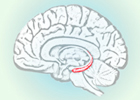
Memory loss is often the result of brain injury and has been linked to damage of the brain structure known as the hippocampus (in red above).
Traumatic brain injury (TBI) is a leading cause of injury-related death and disability in North America. Currently, TBI is seen as "non-progressive"—thus, after injury, it is expected that patients should exhibit steady cognitive improvements and stabilization of brain function. This view is now being challenged by studies that show that progressive loss of brain matter may occur in the chronic stages of injury; furthermore, some patients are known to experience chronic loss of cognitive function as well. Given the possibility that TBI may have long-term risks, new approaches are required to minimize loss of brain function in the chronic stages of injury.
TRI Senior Scientist Dr. Robin Green has been studying this new conceptualization of brain injury and has recently published encouraging findings. Examining whether loss of brain matter could be offset by environments that stimulate high levels of mental, physical and social activity after TBI, Dr. Green's team examined 25 patients with moderate-severe TBI. Those exposed to challenging environments showed reduced loss of brain matter, supporting the notion that "use it or lose it" applies to the chronic stages of recovery of the brain after TBI. Dr. Green states, "These findings, although preliminary, lay the foundation for the development of effective strategies that may dramatically improve long-term outcomes after injury."
This work was supported by the Canadian Institute of Health Research, Physicians Services Incorporated Foundation, the Natural Sciences and Engineering Research Council of Canada and the Ontario Neurotrauma Foundation. R. Green is a Tier 2 Canada Research Chair in Traumatic Brain Injury.
Environmental enrichment may protect against hippocampal atrophy in the chronic stages of traumatic brain injury. Miller LS, Colella B, Mikulis D, Maller J, Green RE. Frontiers in Human Neuroscience. 2013 September 24. [Pubmed abstract]
 TGRI Senior Scientist Dr. Abdallah Daar has been appointed to the Scientific Advisory Board of the United Nations Secretary-General. The Board will strengthen global partnerships for sustainable development to provide political leadership grounded in science.
The Board is composed of internationally renowned scientists—including three Nobel Prize winners—representing various fields of natural, social and human sciences. Dr. Daar is the only Canadian appointed to the 26 member Board.
Congratulations to Dr. Daar for this achievement.