 NRx is UHN's monthly research e-newsletter. Through NRx you can read about ongoing research at our five research institutes, the Princess Margaret Cancer Centre, the Toronto General Research Institute (TGRI), the Toronto Western Research Institute (TWRI), the Toronto Rehabilitation Institute (TRI) and the Techna Institute (Techna).
In this issue you can read about:
- A novel approach for identifying cancer-causing genes
- A new tool for studying SETD7 protein function
- Early detection of kidney disease using magnetic resonance imaging
- Measuring sleep apnea using acoustics
- Regeneration strategies for the injured central nervous system
- CFI's online directory of Canada's cutting-edge research facilities
We hope that you will find NRx informative. If you have feedback or questions, please contact www@uhnresearch.ca.
Christopher J. Paige, PhD, FCAHS
Vice President, Research
University Health Network

A cell's DNA accumulates mutations until a combination of errors in key genes transforms it into a cancer cell.
Cancers arise due to spontaneous changes in DNA, which accumulate over time and cause unrestricted cell growth. The accumulation of these changes makes it challenging to ascertain which errors initiated the cancer. To bypass this problem, researchers normally introduce DNA errors into cells and use the cells as a tool to identify cancer-causing genes. Unfortunately, this strategy has only been successfully achieved using cells from zebrafish and mice. PM Senior Scientist Dr. Rama Khokha and her team have now resolved this issue using several cutting-edge genomic techniques and successfully introduced traceable genome-wide DNA errors into normal human cells.
The study used a novel combination of retroviruses and short DNA sequences (transposons) to insert DNA at random sites across the genome. This rapidly transformed the normal cells into tumour cells with DNA alterations comparable to those found in multiple human cancers. Detailed genomic analyses of these newly generated tumours yielded 80 candidate genes with the potential to drive cancer growth. Importantly, one of the genes found to be defective in at least one in ten tumours has previously been shown to suppress cell growth and is involved in regulating DNA organization.
As Dr. Khokha explains “Our results reveal the potential for using viruses and transposons to rapidly uncover new cancer-causing targets, which will accelerate the global effort to decipher the genes, pathways and networks that drive cancer development and growth.”
This work was supported by the Ontario Institute for Cancer Research, the Canadian Cancer Society Research Institute and The Princess Margaret Cancer Foundation.
Human somatic cell mutagenesis creates genetically tractable sarcomas. Molyneux SD, Waterhouse PD, Shelton D, Shao YW, Watling CM, Tang QL, Harris IS, Dickson BC, Tharmapalan P, Sandve GK, Zhang X, Bailey SD, Berman H, Wunder JS, Iszvak Z, Lupien M, Mak TW, Khokha R. Nature Genetics. 2014 Aug 17. [Pubmed abstract]
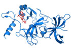
Researchers used a powerful technique known as X-ray crystallography to obtain three-dimensional images of (R)-PFI-2 (red) bound to SETD7 (blue), as shown above. (PDB: 4JLC)
The protein SETD7 is found in different types of cells and is implicated in many physiological processes including metabolism and inflammation. Researchers believe that SETD7 influences these processes by adding specific chemical tags known as methyl groups to other proteins. However, how this activity contributes to the normal functioning of cells is not fully understood by researchers, nor what role the disruption of SETD7 activity plays in disease.
A team lead by Dr. Cheryl Arrowsmith, a Senior Scientist at PM Cancer Centre, in partnership with Pfizer, has synthesized a new chemical compound intended for use by other researchers to shed further light on the biological role of SETD7.
In collaboration with Pfizer, Dr. Arrowsmith’s team screened a library of 150,000 compounds and identified one that inhibited SETD7's activity. This “hit” compound was chemically modified to enhance its potency and facilitate its entry into cells. An atomic-level picture revealed that the modified compound, referred to as (R)-PFI-2, blocked SETD7 activity by attaching directly to it. Researchers then confirmed that (R)-PFI-2 could enter cells, bind tightly and specifically to SETD7 and, as a result, alter the activity of these cells.
“Our collaboration with Pfizer is very exciting. Pfizer is making (R)-PFI-2 freely available to researchers around the world to study the function of SETD7 and identify diseases that it might be able to help treat,” says Dr. Arrowsmith.
This work was supported by the Canadian Institutes of Health Research (CIHR), the Canada Foundation for Innovation (CFI), The Princess Margaret Cancer Foundation and the Structural Genomics Consortium which is a registered charity that receives funds from AbbVie, Boehringer Ingelheim, CFI, CIHR, Genome Canada, the Ontario Genomics Institute, GlaxoSmithKline, Janssen, Lilly Canada, the Novartis Research Foundation, the Ontario Ministry of Economic Development and Innovation, Pfizer, Takeda, and the Wellcome Trust.
(R)-PFI-2 is a potent and selective inhibitor of SETD7 methyltransferase activity in cells. Barsyte-Lovejoy D, Li F, Oudhoff MJ, Tatlock JH, Dong A, Zeng H, Wu H, Freeman SA, Schapira M, Senisterra GA, Kuznetsova E, Marcellus R, Allali-Hassani A, Kennedy S, Lambert JP, Couzens AL, Aman A, Gingras AC, Al-Awar R, Fish PV, Gerstenberger BS, Roberts L, Benn CL, Grimley RL, Braam MJ, Rossi FM, Sudol M, Brown PJ, Bunnage ME, Owen DR, Zaph C, Vedadi M, Arrowsmith CH. Proceedings of the National Academy of Sciences USA. 2014 Aug 18. [Pubmed abstract]
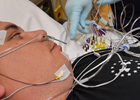
Traditional sleep studies to measure obstructive sleep apnea treatments involve the use of several pieces of equipment and many electrodes.
Obstructive sleep apnea (OSA) is a common condition where the upper airway becomes periodically blocked or collapsed during sleep, causing short pauses in breathing. The underlying mechanisms responsible for this collapse are not fully understood. The accumulation of fluid in the neck appears to play a role—perhaps by increasing the pressure on the airway, thus narrowing it—and research groups are developing treatments to reduce the fluid build-up and prevent OSA and its associated health risks. TRI Scientist Dr. Azadeh Yadollahi and colleagues have developed a convenient and non-invasive technique to measure fluid accumulation in the neck and to monitor the severity of sleep apnea so that the efficacy of these future treatments can be evaluated properly.
Traditional methods for estimating fluid accumulation in the neck include carefully measuring the neck circumference, or using electrodes on the skin to measure the changes in electrical conductivity associated with changes in fluid retention, known as bioelectrical impedance. The TRI team’s innovation was to use the change in breathing sounds and vibrations in the tissue to estimate the fluid volume changes. In a recent study, they were able to demonstrate that sound features captured by a microphone that had been placed on the trachea changed with fluid accumulation in the neck. Moreover, with personalized computer models they could estimate the fluid volume with 98% accuracy.
“This reliable and convenient screening technique has the potential to provide an invaluable tool with which we can estimate neck fluid volume and evaluate potential treatments for OSA,” says Dr. Yadollahi.
This work was supported by the Canadian Institutes of Health Research, Mitacs, the Natural Sciences and Engineering Research Council, the Science Without Borders program between Brazil and Canada, and the Toronto Rehab Foundation.
Acoustic estimation of neck fluid volume. Yadollahi A, Rudzicz F, Mahallati S, Coimbra M, Bradley TD. Annals of Biomedical Engineering. 2014 Aug 8. [Pubmed abstract]
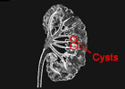
Kidney cysts (circled) are round pouches of fluid that disturb normal functioning of the organ.
Autosomal dominant polycystic kidney disease (ADPKD) is the most common hereditary kidney disease in the world. Although kidney cysts develop normally as part of the natural aging process, the rapid appearance of ADPKD-related cysts eventually leads to organ failure. ADPKD is diagnosed using ultrasonography—an imaging technique employed to visualize and count kidney cysts. Standardized diagnostic criteria using this approach are in place for people over the age of 40. However, ultrasonography is a sub-optimal way to diagnose younger people due to the smaller size of the cysts they develop. This is of particular importance for those who are considered at risk because they have a family history of the disease.
TGRI Senior Scientist Dr. York Pei evaluated two novel imaging techniques for diagnosis of ADPKD in people under the age of 40. He compared high resolution (HR) ultrasound and magnetic resonance imaging (MRI)—both with increased sensitivity in detecting small cysts down to 2-3 mm—with conventional ultrasonography for their ability to diagnose ADPKD. His team found that both imaging tools were more accurate than conventional ultrasonography for early diagnosis as well as exclusion of ADPKD in this young population. Between the two, MRI provided more robust performance than HR-ultrasound in detecting kidney cysts.
Validating the use of MRI to diagnose ADPKD improves our ability to specifically detect early signs of ADPKD in at-risk people under the age of 40, and helps exclude ADPKD in young people who are considering donating a kidney.
This work was supported by the Physicians Services Incorporated Foundation, the National Institutes of Health for the Genetics Core of the Mayo Polycystic Kidney Disease Centre and the Toronto General & Western Hospital Foundation.
Imaging-based diagnosis of autosomal dominant polycystic kidney disease. Pei Y, Hwang YH, Conklin J, Sundsbak JL, Heyer CM, Chan W, Wang K, He N, Rattansingh A, Atri M, Harris PC, Haider MA. Journal of the American Society of Nephrology. 2014 July 29. [Pubmed abstract]
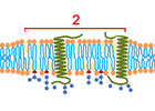
Lipid rafts (region 2 in this schematic of the plasma membrane) compartmentalize cellular processes by influencing membrane fluidity and receptor trafficking.
Injury to the central nervous system (CNS; brain, spinal cord) often results in a permanent loss of function in adults due to the poor capacity of the CNS to regenerate or heal. This is further aggravated by the tendency of brain cells (neurons) to die following injury. A recent study from TWRI Senior Scientists Drs. Philippe Monnier and Charles Tator investigated a transmembrane receptor protein called Neogenin, which is known to control neuron survival or death.
The researchers studied the behaviour of Neogenin in lipid rafts, which are cholesterol-rich regions in the cell membrane where signaling molecules and receptors assemble. In an elegant series of molecular biology experiments, the team showed that Neogenin was recruited to lipid rafts after it interacted with its ligand RGMa. By preventing this interaction with chemical inhibitors or by reducing the amount of membrane cholesterol, they were able to block Neogenin raft localization, promote cell growth and prevent cell death. Blocking Neogenin raft localization also promoted survival and regeneration in the injured adult optic nerve and spinal cord.
“When we lowered cholesterol and disrupted rafts, locomotor function was restored in an experimental model of spinal cord injury,” says Dr. Monnier. “This exciting data presents a potential new strategy to promote regeneration and functional recovery in the injured CNS.”
This study was supported by the Heart & Stroke Foundation of Ontario, the Glaucoma Research Society of Canada, the Canadian Institutes of Health Research and the Toronto General & Western Hospital Foundation.
Modifying lipid rafts promotes regeneration and functional recovery. Tassew NG, Mothe AJ, Shabanzadeh AP, Banerjee P, Koeberle PD, Bremner R, Tator CH, Monnier PP. Cell Reports. 2014 Aug 5 . [Pubmed abstract]

The Canada Foundation for Innovation (CFI) has launched a new online tool, the Research Facilities Navigator, which lists over 360 laboratory profiles from 63 universities, colleges and research hospitals across Canada. The site serves as a quick search point for researchers and businesses to identify facilities and equipment to advance their research and innovation needs.
The research labs listed on the site span five areas: Engineering, Health, Environment, Science, and Social Sciences and Humanities. Examples of labs include University of Waterloo's FELT lab, a public-private partnership that is outfitted with state-of-the-art display technologies to study new ways of interacting with technology; University of Western Ontario's Sustainable Archaeology laboratory, which has imaging equipment to visually reconstruct archaeological sites; and UHN's iDAPT Centre for Rehabilitation Research, which has custom-built immersive environments to recreate challenging environments (eg, the Winter Lab, which simulates snow, ice and wind conditions to safely study fall prevention). UHN's Philip S. Orsino Cell Therapy Facility, Princess Margaret Genomics Centre, Spatio-Temporal Targeting and Amplification of Radiation Response (STTARR) program, Guided Therapeutics (GTx) Program and Electronic Living Laboratory for Interdisciplinary Cancer Survivorship Research (ELLICSR) are also listed on the site.
For more details on these facilities visit the CFI Navigator website.
 UHN's Board of Trustees and the Selection Committee has concluded its international search for a new President and CEO, identifying Dr. Peter Pisters of MD Anderson Cancer Centre.
"As a Canadian, I have always been proud of the health care offered to all citizens," said Dr. Pisters. "To return as President and CEO of UHN is a unique challenge and a privilege. I will do everything in my power to build on the legacy of those who have gone before me."
Dr. Pisters begins his role at UHN on January 1, 2015. He will bring with him a wide range of administrative and leadership experience from his current role at MD Anderson Cancer Center in Houston, Texas, where he is the Vice-President, Regional Care System. He is also an internationally renowned academic surgical oncologist with a specialty focus in sarcoma and gastrointestinal cancers, and an Executive Fellow in the Baldridge Performance Excellence Program of the US National Institute of Standards and Technology. Dr. Pisters is a graduate of The University of Western Ontario (MD) and Harvard University School of Public Health (MSc in Health Care Management).
"UHN's Board of Trustees and the Selection Committee knew how important it was to find someone who understood and was committed to superb clinical care, world-class research and a teaching environment that supports and encourages all members of UHN's community. We also knew that we had to find someone who was completely committed to our efforts to have our patients know that they are partners in their care," said John Mulvihill, Chair of the Board of Trustees.
 UHN's Krembil Discovery Tower (KDT), which is home to one of Canada's most eminent neuroscience, vision and arthritis research programs, has been named one of the 50 most beautiful University Hospitals in North America by the "Best Master of Science in Nursing Degrees" website.
Among the unique architectural features acknowledged by the site were KDT's communal two-storey high collaboration centres, known as Sky Lobbies, which serve as informal areas where research teams can gather together and share their ideas against the backdrop of Toronto's spectacular skyline. The website also noted KDT's sustainability features such as its ample bicycle parking and showers, which promote green transportation and the construction team's recycling initiatives, which enabled 75% of the construction waste to be recycled.
KDT was made possible through a $30 million seed donation from the Krembil family, along with $30 million in grant funding from the Canada Foundation for Innovation. Generous donations by individuals, charities and the Toronto General & Western Hospital Foundation contributed to leveraging additional partner funding. In total, $174 million was raised to make KDT a reality.