
September 2011
Welcome to Net Results EXPRESS
Net Results EXPRESS (NRx) is an award-winning, monthly e-newsletter highlighting medical and scientific breakthroughs, major grants and honours awarded, and other research-related events at UHN. Through NRx you can read about ongoing research at our four research institutes, the Ontario Cancer Institute (OCI), the Toronto General Research Institute (TGRI), the Toronto Western Research Institute (TWRI) and the Toronto Rehabilitation Institute (TRI). We hope you will find this newsletter informative and helpful. If you have feedback or questions, please contact www@uhnresearch.ca. Christopher J. Paige, PhD, FCAHS |
UHN Medical Imaging Tool Licensed to Vexim SA
The software and algorithm, developed by Paul Dufort in UHNs medical imaging group, allows for the recognition and segmentation of highly deformed objects from medical scans taken in a range of contexts. Changes in the object's shape can then be quantified via deformation fields linking multiple scans of the object over time. These are two high-impact and widely known problems in medical image computing which have previously defied general-purpose solutions. This software is a general algorithm that can be tailored to specific applications, with spine injury repair being the focus of the Vexim relationship. The software will continue to be improved and extended, with the next phase of work aimed at incorporating MRI scans to address growing concerns about radiation exposure from CT. |
 UHN has signed a follow-on licensing agreement with Vexim SA, regarding a deformable template registration and segmentation technology developed by UHNs medical imaging group. The agreement, licensed through the Technology Development and Commercialization Office at UHN, provides Vexim (a company based in Toulouse, France) with a robust software algorithm for measuring the efficacy of their SpineJack® implant procedure for repairing vertebral compression fractures. The SpineJack device is a vertebral body implant for the stabilization of vertebral fractures and for restoring height in a minimally invasive, image-guided way. The addition of the UHN software to quantify the effects of this technique provides an important advance in both the planning and the treatment aspects of the SpineJack procedure.
Cancer: Creating a Blueprint of Protein Modifications
Work from the laboratory of OCI Director Dr. Ben Neel has examined the profiles of oxidized PTPs in a number of different human cancer cells. Graduate student Robert Karisch devised a system where all forms of PTPs, oxidized or not, were purified and identified. They identified a number of PTPs that were specifically oxidized in cancer cells and discovered that these modifications could have a number of effects on cellular functions that would contribute to progression of this disease. The identification of oxidized PTPs in this study provides a "blueprint" of the different PTPs that regulate cellular processes important for the development of cancer. "Our results argue strongly that altered oxidation provides an additional level of complexity to the composition of cancer," adds Dr. Neel. Global proteomic assessment of the classical protein-tyrosine phosphatome and "redoxome". Karisch R, Fernandez M, Taylor P, Virtanen C, St-Germain JR, Jin LL, Harris IS, Mori J, Mak TW, Senis YA, Ostman A, Moran MF, Neel BG. Cell. 2011 September 2. [Pubmed abstract] This work was supported by grants from the National Institutes of Health, the Human Frontiers of Science Program, the Canadian Institutes of Health Research, the Canadian Cancer Society Research Institute, the Swedish Research Council, the EU-funded PTPNET Research Training Network, the Ontario Ministry of Health and Long-Term Care and the Princess Margaret Hospital Foundation. |
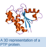 Protein-tyrosine phosphatases (PTPs) are a family of proteins that serve as critical regulators of cell growth. They are highly susceptible to a process called oxidationthis is a modification that can occur when proteins interact with chemically reactive molecules containing oxygen (reactive oxygen species). Oxidation is known to inactivate PTP proteins, which can impact the health and function of cells. Higher levels of reactive oxygen species are linked to a number of diseases, including cancer.
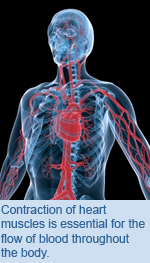
Cardiology: Distinguishing the Mechanisms of Heart Contraction
In their study, Dr. Backx and his colleagues generated mice lacking the specific PDE gene called PDE4D. The hearts of these mice beat more strongly than normal mice while normal mice treated with a drug that inhibit all members of the PDE4 family enzymes also showed elevated Ca2+ and contraction strength. They also showed that PDE4D exerted its effects on cAMP levels and Ca2+ levels by directly interacting with a complex called the SR-Ca2+-ATPase, which controls Ca2+ in cardiac muscle cells. Because levels of cAMP and activity of the SR-Ca2+-ATPase are intensely disrupted in patients with heart failure, Dr Backx proposes, "Our studies demonstrate that selective targeting of PDE4D alone, without affecting other PDEs, could prove helpful in treating heart disease by specifically elevating cAMP levels in cardiac cells." Phosphodiesterase 4D regulates baseline sarcoplasmic reticulum Ca2+ release and cardiac contractility, independently of L-type Ca2+ current. Beca S, Helli PB, Simpson JA, Zhao D, Farman GP, Jones P, Tian X, Wilson LS, Ahmad F, Chen SR, Movsesian MA, Manganiello V, Maurice DH, Conti M, Backx PH. Circulation Research. 2011 September 8. [Pubmed abstract] This work was supported by grants from the Canadian Institutes of Health Research, the Leducq Foundation, the National Institutes of Health and the United States Department of Veterans Affairs Medical Research Funds. |
 Work from the laboratory of TGRI Senior Scientist Dr.
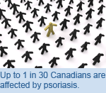
 Psoriatic Arthritis: The Disease within a Disease
The group examined genetic differences in the human leukocyte antigen (HLA) genes in patients with PsA and those with psoriasis without arthritis. HLA is a family of genes that play a role in the immune system and have been previously found to be associated with a number of autoimmune diseases, including Type I diabetes and celiac disease. The gene HLA-B*27 was found to be strongly associated with the development of PsA in patients with psoriasis. Other HLA genes, including C*06, B*38 and B*08, were also found to be associated with the development of PsA. The results of this study provide a clear image of the genetic factors involved in the development of these two diseases. "Patients with psoriasis that are positive for the factors identified in this study may be more susceptible to the development of PsA. These findings may be used to identify patients with psoriasis who could develop PsA early and provide them with appropriate treatment," says Dr. Gladman. Human leukocyte antigen risk alleles for psoriatic arthritis among patients with psoriasis. Eder L, Chandran V, Pellet F, Shanmugarajah S, Rosen CF, Bull SB, Gladman DD. Annals of the Rheumatic Diseases. 2011 September 6. [Pubmed abstract] This work was supported by grants from Krembil Foundation, the Arthritis Society SPARCC National Research Initiative, the Canadian Arthritis Network and the Canadian Institutes of Health Research. |
 A new study from TWRI Senior Scientist and AARC Researcher Dr.
 Nanomedicine: Delivery of Anticancer Drugs
The nanoparticles are composed of a class of proteins, known as self-assembling peptides, which form a structure around ellipticine. The group observed that the drug-linked nanoparticle successfully penetrated lung cancer cells and was safely released into the cell where it could exert its anti-cancer effects. Due to their biocompatible nature, nanoparticles can safely deliver therapeutics without any potential side effects. Self-assembling peptide nanoparticles, in particular, are not toxic and are ideal for biomedical applications, says Dr. Liu. They also can act as a new delivery method for drugs whose chemical properties prevent them from entering cancer cells. Self-assembling peptide based nanoparticles enhance cellular delivery of the hydrophobic anticancer drug ellipticine through caveolae-dependent endocytosis. Bawa R, Fung SY, Shiozaki A, Yang H, Zheng G, Keshavjee S, Liu M. Nanomedicine. 2011 August 31. [Pubmed abstract] This work was supported by grants from the Canadian Institutes of Health Research, the Ministry of Research and Innovation of Ontario, the Uehara Memorial Foundation and the International Society of Heart and Lung Transplantation. |
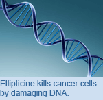 The anticancer drug ellipticine has been shown to kill cancer cells in laboratory conditions, but due to its chemical properties it is unable to effectively target and enter the cells in clinical settings. Nanoparticlesminute molecules with novel propertiescan act as delivery vehicles for anti-cancer drugs. A recent study by TGRI Senior Scientist Dr.
 Cardiology: Reducing Inflammation in Damaged Cardiac Muscle
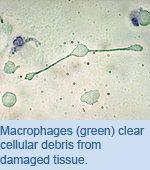
Mice that received an infusion of MSCs following MI were found to have lower levels of pro-inflammatory macrophages and higher levels of anti-inflammatory macrophages in damaged areas of heart muscle. The increase in anti-inflammatory macrophages was concurrent with an increase in IL-10, a factor thought to mediate the switch from pro-inflammation to anti-inflammation. IL-10 is secreted by the infused MSCs, suggesting that they exert control over inflammation in areas of damaged tissue. "Our study warrants further research into investigating the exact role of IL-10 secretion by MSCs in cardiac regeneration," adds Dr. Keating. These results provide increased evidence for the therapeutic use of MSCs following MI. Mesenchymal stromal cells mediate a switch to alternatively activated monocytes/macrophages after acute myocardial infarction. Dayan V, Yannarelli G, Billia F, Filomeno P, Wang XH, Davies JE, Keating A. Basic Research in Cardiology. 2011 September 8. [Pubmed abstract] This work was supported by the Orsino Translational Research Laboratory and the Gloria and Seymour Epstein Chair in Cell Therapy and Transplantation. |
 During a myocardial infarction (MI), more commonly known as a heart attack, muscle tissue in the heart is damaged by oxygen loss and inflammation. Previous studies have shown that the administration of mesenchymal stromal cells (MSCs)cells that are capable of developing into a number of different cell typeswithin the affected heart tissue following MI resulted in decreased injury and improved heart function. A study from TGRI Senior Scientist and Director of the Princess Margaret Hospital Cell Therapy Program Dr.
 Stroke Prevention: Assessing the Effects of Surgical Revascularization
Thirty-nine patients diagnosed with moyamoya vasculopathy, representing 55 brain hemispheres, were assessed through measurements of cerebrovascular reactivity (CVR)increases in blood flow in response to blood pressure stimuliusing blood oxygen level-dependent (BOLD) MRI. CVR measurements preoperatively and 3 months postoperatively revealed that surgery reversed CVR impairment in 52 hemispheres. In a clinical follow-up, 37 of 39 patients had stable or improved functional post-surgery outcomes. "Our data suggests that brain revascularization is safe and effective at reversing the CVR defect," says Dr. Tymianski. "Measurements of CVR with BOLD MRI predict successful arterial opening and clinical response to treatment, making this technique a useful means to evaluate moyamoya patients both preoperatively and postoperatively." Impact of extracranial-intracranial bypass on cerebrovascular reactivity and clinical outcome in patients with symptomatic moyamoya vasculopathy. Han JS, Abou-Hamden A, Mandell DM, Poublanc J, Crawley AP, Fisher JA, Mikulis DJ, Tymianski M. Stroke. 2011 September 8. [Pubmed abstract] This work was supported by the Canada Research Chair in Translational Stroke Research, the Chair Fund of the Neurovascular Therapeutics Program from the University Health Network and a grant from the Ontario Research Fund Brain Consortium. |
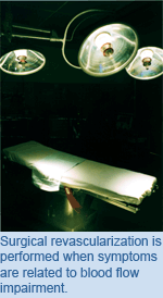 Moyamoya vasculopathy is a progressive disease characterized by the abnormal narrowing of the internal carotid arteriesthese are the major arteries of the head and neck that supply blood to the brainresulting in an increased risk of hemorrhage and stroke. One form of treatment to combat this disease is surgical revascularization, where blood flow is re-established through a reconnection of the cortical arteries or by placing vascularized tissue in direct contact with the brain to promote new blood vessel growth. A study by TWRI Senior Scientists Dr.
![]()
 Dr. Anthony Lang, TWRI Senior Scientist and Director of the Morton and Gloria Shulman Movement Disorders Centre, has been named a Fellow of the Royal Society of Canada (RSC). Dr. Lang is recognized as one of Canadas leading experts in Parkinsons disease and was inducted into the Society for his clinical and research contributions to this disease. In December 2010 he was appointed as an Officer in the Order of Canada and earlier this year was also elected as a Fellow of the Canadian Academy of Health Sciences. Between 2001 and 2010, Dr. Lang was the worlds most cited author in the field of Parkinsons disease research.
The RSC is a Canadian institution devoted to recognizing excellence in learning and research as well as accomplishments in the arts, humanities and sciences. It consists of approximately 2000 Fellows, a collection of distinguished scholars, artists and scientists and serves as Canadas national academy. The objective of the RSC is to recognize academic excellence, to advise governments and organizations, and to promote Canadian culture.
Congratulations Dr. Lang!
 UHN congratulates TGRI Senior Scientist Dr. Ren-Ke Li for his recent election as a Fellow of the Canadian Academy of Health Sciences (CAHS). Dr. Li is recognized for his contributions to the field of cardiac regeneration and repair. His work has focused on the clinical applications of transplantation of healthy cardiac cells into injured hearts, as well as the use of tissue engineering to create cardiac tissue for the repair of injured hearts to improve heart function.
The Academy was formed in 2004 to serve as a source of expert advice on issues related to the health of Canadians. Fellows of CAHS are elected on the basis of their leadership, creativity and commitment to the advancement of the academic health sciences. Dr. Li joins other UHN researchers like Drs. Christopher Paige, Abdallah Daar, Peter Singer and Charles Tator in the Academy.
 UHN welcomes Dr. Daniel Winer to the Toronto General Research Institute as a Scientist in the Division of Cellular and Molecular Biology and a Pathologist in the Department of Pathology. Dr. Winer joins UHN from Stanford University in California where he spent a number of years studying the development of diabetes in Dr. Edgar Englemans lab.
Dr. Winers research at TGRI will focus on better understanding how the immune system influences physiological processes involved in disease. His studies will expand upon his recent findings that the adaptive immune systemincluding T cells, B cells and the antibodies they produceplay a significant and active role in regulating the process of insulin resistance. Dr. Winers lab will investigate immune-mediated mechanisms in obesity and diabetes with the aim of translating their findings to help the many people afflicted by these diseases.
The CoEN Initiative brings together leading international laboratories to undertake innovative research that will increase our understanding of how neurodegenerative diseases are triggered and progress, accelerating the development of new approaches to treatment.
This grant was one out of eight applications that received funding in this initiative.
Feedback
Net Results EXPRESS is brought to you by UHN Research Communications. We hope you have enjoyed receiving this message. If you have any feedback, please email www@uhnresearch.ca.
To access archived issues of Net Results EXPRESS, visit uhnresearch.ca/news/netresultsexpress
Some images adapted from the image archives of stock.xchng.ca.