

An extensive network of branching nerves enables the face to feel a wide range of sensations.
However, when these nerves become damaged or irritated—as is the case in a condition known as trigeminal neuralgia—they can become a source of chronic and excruciating bursts of pain. For individuals with the condition, even simple actions like chewing food can trigger debilitating pain.
Krembil Scientist and neurosurgeon Dr. Mojgan Hodaie, together with her research team, has mapped out key differences in the nerves of people suffering from trigeminal neuralgia to better understand the disease.
“This is the first in-depth survey of microarchitectural abnormalities in nerves associated with trigeminal neuralgia,” says Dr. Hodaie. “We traced the entire nerve pathway—from peripheral segments of the trigeminal nerve to where its branches gather, all the way to the brain.”
Using magnetic resonance imaging (MRI), the research team collected detailed images of the tissues in the faces of patients with or without the condition. To study whether the disease causes structural differences in the trigeminal nerve or its connections, the researchers used programs to automatically generate 3D models of the nerve pathways from the MRI data for all the subjects, with little human intervention.
Because these 3D models were highly complicated, Dr. Hodaie’s team applied machine learning algorithms—a form of artificial intelligence—to pick out the hidden differences. These algorithms enabled the team’s computer to learn which features corresponded to patients with trigeminal neuralgia and pinpoint where the differences occurred.
The team’s analysis revealed abnormalities in the nerves of individuals with trigeminal neuralgia that could be used to distinguish them from healthy subjects with more than 80% accuracy. For patients with pain in only one side of the face, these abnormalities could likewise be used to distinguish the unaffected and affected sides.
The latter analysis also revealed a peculiarity: patients with pain in only one side of their face still had some abnormalities on both sides. This intriguing result, along with other findings, lays the foundation for future research and the development of therapeutic strategies to target the observed differences.
This work was supported by the Multiple Sclerosis Society of Canada and Toronto General & Western Hospital Foundation.
Chen DQ, Zhong J, Chu PPW, Fei Li CM, Hodaie, M. Trigeminal Neuralgia Diffusivities using Gaussian Process Classification and Merged Group Tractography. Pain. 2020 Jul 21. doi: 10.1097/j.pain.0000000000002023.
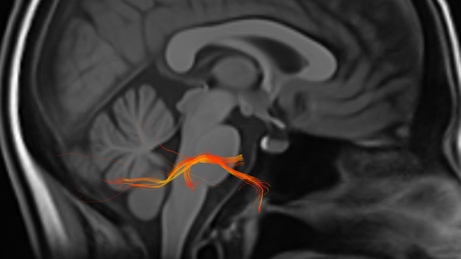
A 3D model of the right-side trigeminal nerve (orange) overlaying an MRI scan of the head. The trigeminal nerve branches into three divisions, which supply nerves to areas of the face such as the jaw, cheeks, nose, sinuses, eyelids, brow and forehead.